Near infrared laser scanning confocal microscopic imaging system

A confocal microscopy and laser scanning technology, applied in microscopes, optics, optical components, etc., can solve problems such as large scattering and absorption of biological tissue, biological tissue damage, and unfavorable imaging depth, and achieve deep imaging depth and biological tissue damage. Small, flexible selection of effects

Inactive Publication Date: 2017-04-19
ZHEJIANG UNIV
View PDF14 Cites 11 Cited by
  • Summary
  • Abstract
  • Description
  • Claims
  • Application Information

AI Technical Summary

Problems solved by technology

This laser scanning confocal microscopy imaging system based on excitation in the visible light band and fluorescence reception and imaging in the visible light band has limited selection of excitation light wavelengths, and they are all in the visible light band. There is

Method used


Image

  • Near infrared laser scanning confocal microscopic imaging system

Examples


Embodiment Construction

[0012] The present invention will be further described below in conjunction with accompanying drawing.

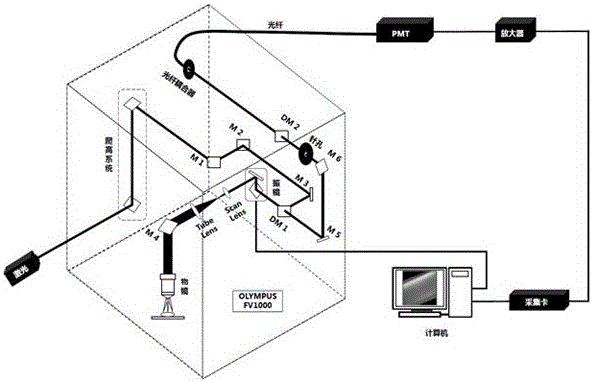
[0013] Such as figure 1 As shown, the near-infrared laser scanning confocal microscopy imaging system based on Olympus FV1000 includes near-infrared laser light source, optical path climbing system, Olympus FV1000 laser scanning confocal upright microscope, dichroic mirror, optical fiber Coupler, near-infrared response PMT, signal amplifier, acquisition card, computer, etc.

[0014] First, the external near-infrared laser is introduced into the Olympus FV1000 laser scanning confocal microscopy imaging system through an optical path climbing system. The optical path is reflected by a long pass (short reflection) dichroic mirror and enters the galvanometer scanning unit. Then through the beam expander system composed of scanning lens (Scan lens) and lens tube lens (Tube lens), the outgoing light is reflected by a mirror, converged by the objective lens, and excites the near-...


PUM


Abstract

The invention discloses a near infrared laser scanning confocal microscopic imaging system. A near infrared laser not contained in an Olympus FV1000 laser scanning confocal microscope is selected as a light source, a near infrared fluorescent probe in a biological specimen is stimulated, near infrared fluorescence is led out by optical fibers and is received by PMT responding to the near infrared light and imaging is preformed. Compared with a traditional laser scanning confocal microscopic imaging system performing stimulation and receiving in visible light bands, the near infrared laser scanning confocal microscopic imaging system has the advantages of smaller biological tissue damage, deeper imaging depth, stable work and reliable performance.

Description

technical field [0001] The invention belongs to the field of microscopic imaging of applied optics, and is a near-infrared laser scanning confocal microscopic imaging system modified on the basis of Olympus commercial laser scanning confocal microscope. Background technique [0002] Laser scanning confocal microscope (LSCM for short) is a new type of high-precision microscopic imaging system that combines an optical microscope, a laser light source, a scanning unit, a photodetector, a data acquisition unit and a computer unit. It is based on the traditional fluorescence microscopy imaging system, using laser as the light source, the laser beam converges on the specimen through the objective lens, and forms a small light spot on the focal plane, and the fluorescence excited by the focal point and the nearby sample area is captured by the objective lens Collect and return to the beam splitter composed of dichroic mirrors along the original irradiation light path. The beam spl...

Claims


Application Information

Patent Timeline
IPC IPC(8): G01N21/64G02B21/00
Inventor 钱骏孙超伟
Owner ZHEJIANG UNIV
Why Eureka
Try Eureka