Separable capsule

A separate and capsule-based technology, which is applied in medical science, diagnosis, endoscopy, etc., can solve the problems of occupying the space of the capsule and the inability to detect multiple markers at the same time, and achieve the effect of convenient use, high accuracy, and simple operation

Active Publication Date: 2017-02-01
CHONGQING UNIV
View PDF5 Cites 4 Cited by
  • Summary
  • Abstract
  • Description
  • Claims
  • Application Information

AI Technical Summary

Problems solved by technology

However, due to the limitation of the space at the detection end, the detection marker of the capsule system is only one kind of gastric occult blood, and it cannot detect multiple...

Method used


Image

  • Separable capsule
  • Separable capsule
  • Separable capsule

Examples


Embodiment 1

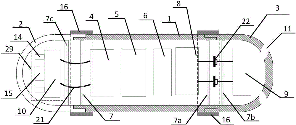
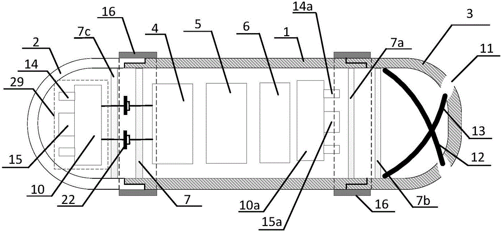
[0029] Such as figure 2 As shown, a detachable capsule includes a main cabin 1, an auxiliary cabin one 2 and an auxiliary cabin two 3, and the main cabin 1 includes a wireless transmitting module 4, a micro battery 5, a power control module 6, a signal acquisition control circuit 8, and a signal acquisition control circuit 8. Acquisition control circuit 8 includes LED lamp group 14a, CMOS sensor 15a, microcontroller 10a; In the auxiliary cabin one 2 is an image acquisition circuit 29, including LED lamp group 14, CMOS sensor 15, microcontroller 10; In the auxiliary cabin two 3 Contains hemoglobin detection test paper 12 and carcinoembryonic antigen detection test paper 13, two through holes 11 are provided, the detection ends of hemoglobin detection test paper 12 and carcinoembryonic antigen detection test paper 13 are pasted on the two through holes 11, and the other end is pasted on the auxiliary cabin Two 3 on the inner wall. The first auxiliary cabin 2 and the second aux...

Embodiment 2

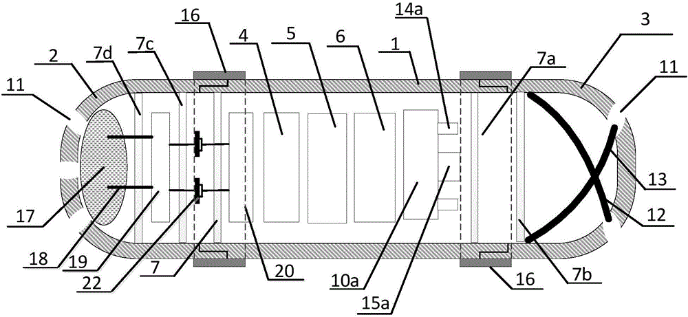
[0037] Such as image 3 Shown, a kind of detachable capsule comprises main cabin 1, auxiliary cabin one 2 and auxiliary cabin two 3, and present embodiment two is compared with embodiment one, and the structure of auxiliary cabin two 3 is exactly the same as the realization method, and main cabin In 1, a signal acquisition control circuit 20 is added; the structure of the auxiliary cabin 1 2 is different from the implementation method; the auxiliary cabin 1 2 and the auxiliary cabin 2 3 are connected and fixed with the main cabin 1 through a degradable fixing ring 16, which is degradable After the fixing ring 16 is degraded, the first auxiliary cabin 2 and the second auxiliary cabin 3 are separated from the main cabin 1 .

[0038] Secondary cabin one 2 of present embodiment two comprises absorbent cotton 17, pH detection probe 18, pH detection sensor 19 and baffle plate 7d, and baffle plate 7d is transparent or opaque, and pH detection sensor 19 selects antimony electrode, pH ...


PUM

PropertyMeasurementUnit
Lengthaaaaaaaaaa
Diameteraaaaaaaaaa

Abstract

The invention provides a separable capsule for detecting a disease marker in gastric fluid and acquiring a real image of the stomach. The separable capsule comprises a main cabin, an auxiliary cabin I and an auxiliary cabin II, wherein the main cabin comprises a wireless emission module, a micro battery, a power control module and a signal acquisition and control circuit; the auxiliary cabin I comprises an image acquisition image or a gastric fluid detection sensor; the auxiliary cabin II comprises an image acquisition circuit or a gastric fluid detection sensor; each image acquisition circuit comprises an LED lamp group, a CMOS sensor and a micro controller; the auxiliary cabin I and the auxiliary cabin II are connected and fixed with the main cabin through degradable fixing rings; after the degradable fixing rings are degraded, the auxiliary cabin I and the auxiliary cabin II are separated from the main cabin. The separable capsule provided by the invention can take a picture of a gastrointestinal wall and can also detect various disease markers; after the detection is completed, the two auxiliary cabins of the capsule are separated from the main cabin, so that the total size of the capsule is decreased, and the risk that the capsule cannot be discharged after entering a human body is effectively reduced.

Description

technical field [0001] The invention relates to a medical detection device, in particular to a detachable capsule for detecting gastrointestinal diseases by swallowing the capsule. Background technique [0002] In clinical medicine, the diagnosis of gastric disease mostly begins with the detection of gastric juice. At present, the main method of extracting gastric juice is to use a gastroscope, inserting an optical microscope from the mouth into the stomach for liquid extraction. The patient suffers a lot. The pain was unbearable, and the gastroscope can only extract gastric juice, and other inspection operations are carried out outside the body. [0003] Patent CN02268048 proposes a gastric juice detector, in which the detection test paper is bundled into a test paper ball with a lead wire, the test paper ball is wrapped in a soluble envelope, and the end of the lead wire is placed outside the soluble envelope. Although it is convenient for patients to use, on the one hand...

Claims


Application Information

Patent Timeline
IPC IPC(8): A61B1/04A61B1/06A61B5/00
CPCA61B1/041A61B1/0684A61B5/4283
Inventor 乔盼盼刘洪英皮喜田
Owner CHONGQING UNIV
Why Eureka
Try Eureka