
Method for isolated culture of umbilical vein endothelial cell

A vascular endothelial, separation and culture technology, applied in the direction of vascular endothelial cells, tissue culture, animal cells, etc., can solve the problems of easy contamination by other cells, large amount of enzymes, leakage, etc., to achieve high cell adhesion efficiency and less pollution , the effect of simple operation

Inactive Publication Date: 2016-08-31
SHANDONG QILU STEM CELL ENG
View PDF3 Cites 1 Cited by
  • Summary
  • Abstract
  • Description
  • Claims
  • Application Information

AI Technical Summary

Problems solved by technology

[0007] In order to solve the above-mentioned problems in the traditional umbilical vein endothelial cell perfusion digestion method in the prior art, which are difficult for a single person to operate, easy to cause leakage, easy to be polluted by other cells, and large amount of enzymes, the inventors made comprehensive considerations, Developed a method for isolating and culturing umbilical vein endothelial cells that is suitable for single-person operation, is relatively simple to operate, is not easily contaminated, and requires a small amount of enzymes

Method used


Image

  • Method for isolated culture of umbilical vein endothelial cell
  • Method for isolated culture of umbilical vein endothelial cell
  • Method for isolated culture of umbilical vein endothelial cell

Examples


Embodiment 1

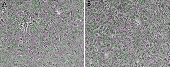
[0031] Implementation Example 1 Carry out the extraction of umbilical vein endothelial cells according to this scheme

[0032] This example uses neonatal umbilical cords with maternal consent as the source of umbilical vein endothelial cells.

[0033] The reagents used in this experiment are as follows: type II collagenase (Sigma), α-MEM medium (Hyclone, Cat.No.SH30265.01B), vascular endothelial growth factor VEGF (Peprotech), fibroblast growth factor FGF (Peprotech ), fetal bovine serum FBS (Gibco), trypsin (Hyclone).

[0034] (1) Wash the neonatal umbilical cord (about 10 cm) three times in normal saline containing 1% double antibody to remove the blood stains on the surface, use sterile ophthalmic scissors to cut the umbilical cord into a small mouth about 0.5 cm along the umbilical vein, and then use sterile Remove the umbilical cord along the longitudinal direction of the umbilical vein with sterile dressing forceps, so that the umbilical vein is completely exposed, and ...

Embodiment 2

[0038] Implementation Example 2 Extraction of Umbilical Vein Endothelial Cells by Collagenase Perfusion

[0039] The reagents used in this experiment are the same as in Example 1.

[0040]The separation of umbilical vein endothelial cells was carried out according to the traditional collagenase perfusion method. Since the method is relatively mature, it will not be repeated here. During the digestion process, the type II collagenase with a mass volume concentration of 0.05% was used to digest for 25 min, and after the cells were collected, 5×10 4 / well into a 6-well plate, placed in 37 ° C, 5% CO 2 After 24 hours, the medium was changed to remove floating red blood cells. After 3 days, the medium was replaced with medium B. Cells were subcultured after the degree of cell confluence reached 80%, and medium B was used in the subsequent culture process. The final concentration of fetal bovine serum FBS in medium A was 20%, the final concentrations of basic fibroblast growth fac...

Embodiment 3

[0043] Compared with Example 1, in the step (3) of this example, type II collagenase was used at a mass volume concentration of 0.1% for 10 cm blood vessels, and the digestion time was 15 minutes, and the rest was the same as that of Example 1.

[0044] In embodiment example 3, 10cm umbilical cord obtains initial cell number and is 1.8 * 10 5 , it takes 144h for the cells to grow to 80% confluence.


PUM


Abstract

The invention relates to the field of isolated culture of adult cells in the technical field of biology, in particular to a method for isolated culture of umbilical vein endothelial cells. The method comprises the following steps: completely stripping an umbilical vein from wharton jelly; longitudinally cutting off the umbilical vein along one side so as to change the umbilical vein into a rectangular blood vessel wall surface from a tube shape and rinsing; adding II-type collagenase for digestion; and taking out the blood vessel and rinsing after digestion, mixing rinsing liquid with enzymatic hydrolysate, centrifuging, removing supernatant and carrying out culture by using a culture medium. The cell separation method is simple to operate, and can be independently carried out by a single person; red cell contamination is relatively low; liquid exchange can be carried out 72 hours after cell inoculation, so that the cell adhesion efficiency is relatively high. A large amount of endothelial cells obtained by primary generation are obtained; CD31 positive expression of P3 cells can reach 95% or above; preparation of umbilical cord mesenchymal stem cells is not affected when the endothelial cells are separately prepared; and the experiment material can be fully utilized.

Description

technical field [0001] The invention relates to the field of separation and culture of adult cells in the field of biotechnology, in particular to a method for separation and culture of umbilical vein endothelial cells. Background technique [0002] Blood vessels are the main body of the human body's internal circulatory system, and the inner surface of the entire blood vessel is covered by a layer of endothelial cells. Because endothelial cells can directly contact with blood, they can play an important role in blood pressure regulation, coagulation and fibrinolysis, adhesion and migration of inflammatory cells, and angiogenesis and other physiological and pathological processes through various channels. [0003] In cell research, vascular endothelial cells are commonly used cells for in vitro experiments, and most studies using human-derived vascular endothelial cells are carried out using human umbilical vein endothelial cells (HUVEC) as materials. . The reason why the ...

Claims


Application Information

Patent Timeline
Patent Type & Authority Applications(China)
IPC IPC(8): C12N5/071
CPCC12N5/069C12N2501/115C12N2501/165
Inventor 刘东曲廷瑜张秀涛
Owner SHANDONG QILU STEM CELL ENG
Learn More