Fast fan-beam geometric phase contrast CT imaging device and method

A phase contrast and CT imaging technology, applied in the field of X-ray imaging, can solve the problems of long phase contrast CT imaging time, limited practical application, and high dose

Inactive Publication Date: 2016-06-15
CHINA HEFEI TAIHE OPTOELECTRONICS TECH
View PDF5 Cites 9 Cited by
  • Summary
  • Abstract
  • Description
  • Claims
  • Application Information

AI Technical Summary

Problems solved by technology

The traditional grating phase-contrast imaging method uses the phase-stepping method to complete information separation and extraction, but the phase-stepping method needs to move the grating laterally, which is incompatible with the CT scanning method, which leads to the disadvantages of long phase-contrast CT imaging time and high dose, and cannot be used for the heart. isokinetic tissue imaging
In 2010, researcher Zhu Peiping published a fast grating phase contrast imaging method (PNAS107, 13576–13581, 2010) in the Proceedings of the National Academy of Sciences of the United States (PNAS107, 13576–13581, 2010), which is the front and back projection method. Applied to parallel beam illumination geometry, usually synchrotron X-ray sources, which greatly limits its practical application

Method used


Image

  • Fast fan-beam geometric phase contrast CT imaging device and method
  • Fast fan-beam geometric phase contrast CT imaging device and method
  • Fast fan-beam geometric phase contrast CT imaging device and method

Examples


Embodiment Construction

[0040] In the hard X-ray illumination of grating phase contrast imaging, the absorption grating in the imaging system requires a large thickness, resulting in a large aspect ratio. In fan-beam geometric illumination, a large aspect ratio flat grating will block rays with large incident angles, which severely restricts the imaging field of view.

[0041] Such as figure 1 As shown, the fast fan beam geometric phase contrast imaging device of the present invention includes an X light source S and a phase grating G 1 , Sample platform P, analysis grating G 2 And detector D. Phase grating G 1 , Analysis grating G 2 Both detector D and detector D are cylindrical elements, and their radius of curvature is equal to the distance between it and the light source.

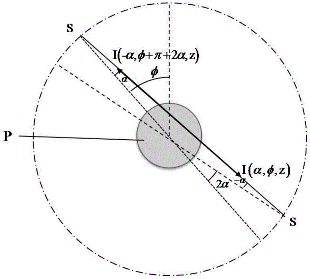
[0042] The fast fan beam geometric phase contrast imaging method of the present invention can pass figure 2 The process shown is completed: first, without placing the object, analyze the grating G along η cycles 2 Moving analysis...


PUM


Abstract

The invention discloses a fast fan-beam geometric phase contrast CT imaging device and method. The phase contrast CT imaging device includes an X light source, a phase grating, a sample platform, an analysis grating and a detector. The invention adopts a cylindrical optical element, Large field of view imaging can be realized under the illumination of conventional X light sources; the invention uses cylindrical optical elements that match the geometry of light to avoid the problem of rays being blocked at large incident angles, greatly improving the imaging field of view, and at the same time combining the idea of ​​square projection, In the traditional CT scanning mode, the phase information of the object is successfully extracted, and the fast phase contrast CT imaging is completed.

Description

Technical field [0001] The invention relates to the technical field of X-ray imaging, in particular to a fast fan beam geometric phase contrast CT imaging device and method. Background technique [0002] Since Roentgen discovered X-rays, X-rays have been widely used in medical imaging, industrial non-destructive testing and other fields. However, traditional X-ray absorption imaging is not ideal for imaging soft tissues with low atomic coefficients. Under X-ray irradiation, the refractive index of an object can be expressed as a complex number as n=1-δ-iβ, where δ is the decrease in the real part of the refractive index and β is the imaginary part of the refractive index. As the atomic coefficient decreases, δ and β decrease, but in the hard X-ray band, the decrease in the real part of the refractive index δ of the soft tissue with low atomic coefficient is about 1000 times the imaginary part β. Therefore, it is theoretically possible to obtain an image with higher contrast than...

Claims


Application Information

Patent Timeline
IPC IPC(8): G01N23/04
CPCG01N23/04
Inventor 吴朝颜天信汪洪波张猛
Owner CHINA HEFEI TAIHE OPTOELECTRONICS TECH
Why Eureka
Try Eureka