Eureka AIR delivers breakthrough ideas for toughest innovation challenges, trusted by R&D personnel around the world.

Method for detecting 5-methylcytosines in nucleic acid

A technology of methylcytosine, nucleic acid, applied in the field of nucleic acid chemistry

Inactive Publication Date: 2015-11-25
WUHAN UNIV
View PDF1 Cites 0 Cited by
  • Summary
  • Abstract
  • Description
  • Claims
  • Application Information

AI Technical Summary

Problems solved by technology

The existing methylation detection methods include liquid chromatography mass spectrometry and mass spectrometry, and chemical methods such as sodium bisulfite method, sodium periodate method, etc., but everything has shortcomings, and these methods still need to be perfected to improve the detection accuracy

Method used


Image

  • Method for detecting 5-methylcytosines in nucleic acid
  • Method for detecting 5-methylcytosines in nucleic acid
  • Method for detecting 5-methylcytosines in nucleic acid

Examples


Embodiment 1

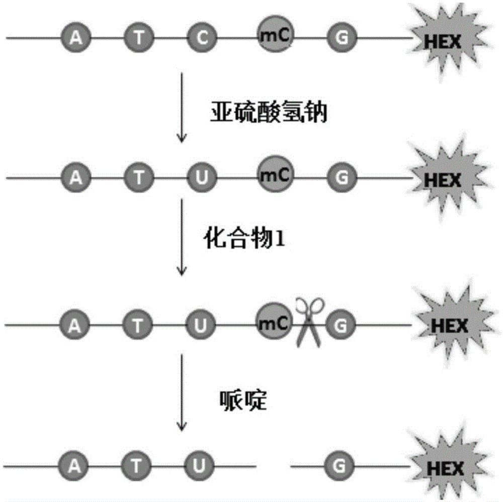
[0023] Example 1: Sodium bisulfite treatment of nucleic acid strands containing 5-methylcytosine

[0024] Pipette 2 μL of 5-methylcytosine-containing DNA (10 μM) labeled with the fluorescent substance HEX (Chinese name is 5-hexachlorofluorescein phosphoramidate) at the 3’ end, add 48 μL of water, and then add 5.5 μL of freshly prepared NaOH solution (3M), incubate in a water bath at 42°C for 0.5 hours, add 30 μL of hydroquinone (10 mM) after the completion of the water bath incubation, and then add 520 μL of NaHSO 3 solution (3.6M, pH=5.0) and 200 μL of paraffin oil, and incubated in a water bath at 50° C. for 16 hours in a dark place, to change cytosine on DNA to uracil. After the reaction, use a desalting column to desalt and purify the DNA.

Embodiment 2

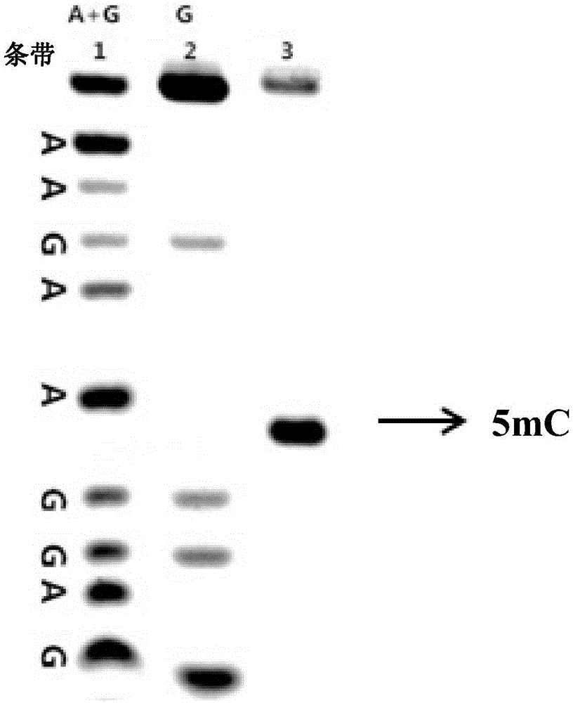
[0025] Example 2: DNA treated with sodium bisulfite was treated with compound 1 and volume fraction of 10% piperidine

[0026] Take 4 μL of 5-methylcytosine-containing DNA (10 μM) treated with sodium bisulfite, add 10 μL of acetonitrile, 2 μL of Tris-HCl solution (1M, pH=5.0), 2 μL of water and 2 μL of compound 1 ( 20mM), heat treatment at 50°C for 10 minutes, 5-methylcytosine on the DNA chain specifically binds to compound 1; then precipitated with ice ethanol, centrifuged, spin-dried the solvent, and then added piperidine with a volume fraction of 10% at 90°C for 0.5 h, the position-specific cleavage of the 5-methylcytosine bound to compound 1; then ice-ethanol precipitation, centrifugation, and spin-drying of the solvent. Load the sample to the electrophoresis tank, and observe the gel results after 2 hours. By comparing with the control of the A+G sequence and the control of the G sequence, the position and number of the DNA bands in the polyacrylamide denaturing gel can b...


PUM


Abstract

The invention provides a method for detecting 5-methylcytosines in nucleic acid. The method includes the concrete steps that first, a nucleic acid chain with a fluorescent label is treated with sodium hydrogen sulfite, all cytosines on the nucleic acid chain will change into uracils, but the 5-methylcytosines are not influenced; then, a compound 1 is specifically reacted with the 5-methylcytosines, but is not reacted with the uracils, thymines, guanines and adenines; due to the fact that thermal piperidines can rupture damaged basic group portions in the nucleic acid, the positions and number of the 5-methylcytosines can be clearly seen through a nucleic acid strip of denatured polyacrylamide gel after the nucleic acid chain reacted with the compound 1 is treated with the thermal piperidines, wherein the structural formula of the compound 1 is shown in the specifications.

Description

technical field [0001] The invention belongs to the field of nucleic acid chemistry and relates to a chemical detection method for 5-methylcytosine in nucleic acid. Background technique [0002] With the rapid development of sequencing technology and the discovery of more and more gene sequences and their functions, human beings are eager to understand various life processes. Genes support the basic structure and functions of life, are the main substance of heredity and variation, and contain all the information of life's gestation, growth, aging, apoptosis and other processes. Epigenetics is an important branch of genetics, which studies the changes in gene expression levels caused by non-gene sequence changes, such as nucleic acid methylation and chromatin conformation changes. People are slowly realizing the importance of epigenetics. [0003] Nucleic acid methylation is known as the epigenetic marker of "gene silencing". Nucleic acid methylation is closely related to ...

Claims


Application Information

Patent Timeline
IPC IPC(8): C12Q1/68
CPCG01N27/447
Inventor 周翔王雅芬毛伍祥刘朝兴
Owner WUHAN UNIV
Why Eureka
Eureka Blog
Learn More