
Cell co-culture micro-fluidic chip and application thereof

A microfluidic chip and co-cultivation technology, applied in the field of biomedicine, can solve problems such as difficult to accurately simulate the real situation in the body, uneven distribution of cells, difficult operation, etc., to achieve wide application value, strong parallel culture ability, and equipment simple effect

Inactive Publication Date: 2015-05-13
THE NAT CENT FOR NANOSCI & TECH NCNST OF CHINA
View PDF0 Cites 11 Cited by
  • Summary
  • Abstract
  • Description
  • Claims
  • Application Information

AI Technical Summary

Problems solved by technology

This traditional culture device has many disadvantages: expensive, uneven distribution of cells, difficulty in accurately simulating the real situation in the body, difficult operation, etc.
However, the use of similar compartmentalized microfluidic chips to simultaneously complete the implantation and co-culture of nerve-cancer cells and study their interaction, as well as further screening of anti-tumor drugs, is still in a blank stage in the current application field.

Method used


Image

  • Cell co-culture micro-fluidic chip and application thereof
  • Cell co-culture micro-fluidic chip and application thereof
  • Cell co-culture micro-fluidic chip and application thereof

Examples


Embodiment 1

[0044] The fabrication method of the cell co-culture microfluidic chip is as follows:

[0045] 1) Coating of the petri dish: add 1 mL of 0.1 mg / mL polylysine (PDL) to a clean petri dish (Corning, 35 mm in diameter) and incubate at 37°C for 1-2 hours to obtain a surface coated with The surface of the petri dish of PLD was washed 3 times with sterile water to remove unadsorbed molecules, and dried in a sterile operating table for later use.

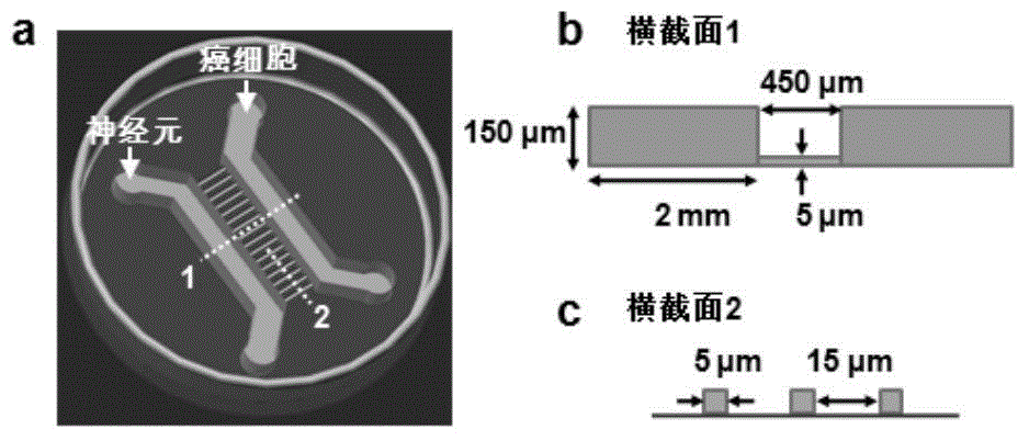
[0046] 2) Fabrication of the SU-8 photolithography mold: a convex microstructure unit with a double-layer structure was prepared on the silicon wafer using multiple photolithography techniques. First, use the drawing software autoCAD to design the required double-layer overlay structure, which includes: linear convex lines located at the left and right positions, and a micro-convex line array connecting the left and right convex lines. The length of the two left and right convex lines is 2 cm, the width is 2 mm, and the height is 150 μm. T...

Embodiment 2

[0052] Using the cell co-cultivation microfluidic chip of the present invention to carry out cell co-cultivation, the specific steps are as follows:

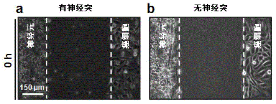
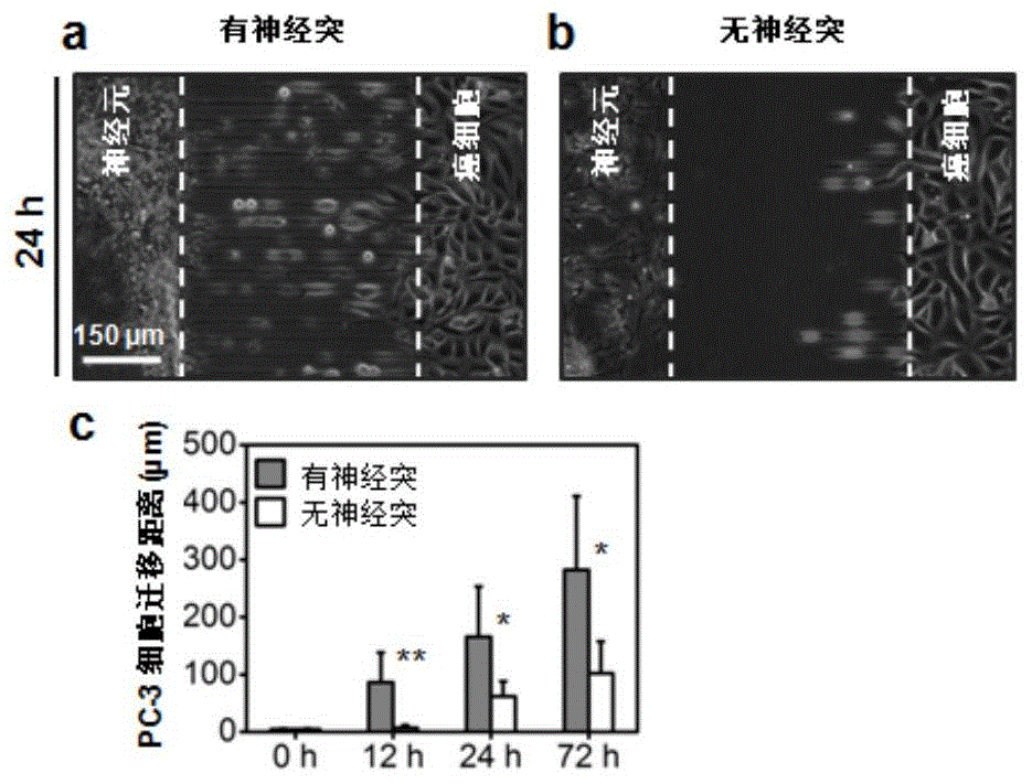
[0053] 1) Prepare a suspension solution of DRG neurons with a cell density of 6 x 10 7 cells / ml, then pass the DRG neurons into the left lumen, add the medium in the opposite channel as a balance, and soak the chip with the medium. Culture medium is Basal medium + 2% B27 complement + 1% PS double antibody. Cultured in a constant temperature incubator for 30-60 minutes, the neuron cells adhered to the surface of the culture dish in the right channel; after 72-96 hours, due to the height restriction, the neuron cell body was restricted to grow in the neuron channel, while the neuron cells The protrusion grows and reaches the opposite channel through the dimpled channel.

[0054] 2) Prepare suspension solutions of different cancer cells (human prostate cancer cell PC-3, human pancreatic cancer cell Panc-1, or human breast cance...


PUM

PropertyMeasurementUnit
Lengthaaaaaaaaaa
Widthaaaaaaaaaa
Heightaaaaaaaaaa

Abstract

The invention relates to a cell co-culture micro-fluidic chip and application thereof. The cell co-culture micro-fluidic chip comprises culture channels and a microchannel, wherein the culture channels are connected through the microchannel; the height of the culture channels is 100-300 microns; the height of the microchannel is less than or equal to 5 microns. The micro-fluidic chip disclosed by the invention is used for constructing a nerve microenvironment of a tumor, so as to provide a platform for studying the tumor microenvironment and basic research on nerve-cancer interaction, further can be used for pharmaceutical detection based on the action between the cells to screen the antitumor effect of the related nerve drugs, and provides a novel approach for analysis to discover the antitumor drugs.

Description

technical field [0001] The invention relates to the field of biomedicine, in particular to a microfluidic chip and its application, in particular to a cell co-cultivation microfluidic chip and its application. Background technique [0002] The tumor microenvironment plays an important role in tumor formation, development, metastasis and recurrence. The nervous system is a common feature of the tissue microenvironment, but its role in tumor growth and progression is still poorly understood. High-density nerve growth in and around tumors has been reported to help cancer grow and spread in animal models. Previous research has also shown that cancer cells sometimes migrate along nerves, but exactly how this works is not well understood. Therefore, establishing an in vitro model of the neural microenvironment of tumors will not only help to elucidate the role of nerves in cancer growth and metastasis, but also may lead us to further develop new strategies for cancer treatment. ...

Claims


Application Information

Patent Timeline
IPC IPC(8): C12M3/00C12N5/09C12N5/079C12N5/0793C12Q1/02
Inventor 蒋兴宇雷祎凤郑文富
Owner THE NAT CENT FOR NANOSCI & TECH NCNST OF CHINA
Learn More