Rare cell detection chip and application thereof
A rare cell and detection chip technology, applied in animal cells, tumor/cancer cells, vertebrate cells, etc., can solve the problems of poor selectivity and sensitivity, achieve the effect of enhancing selectivity or other characteristics and reducing additional pressure
- Summary
- Abstract
- Description
- Claims
- Application Information
AI Technical Summary
Problems solved by technology
Method used
Image
Examples
Embodiment 1
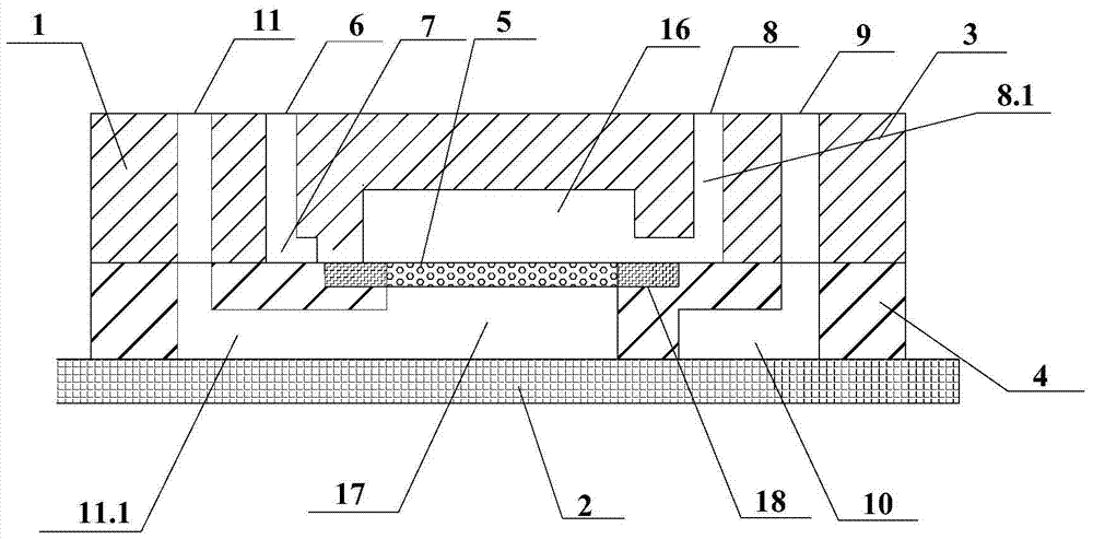
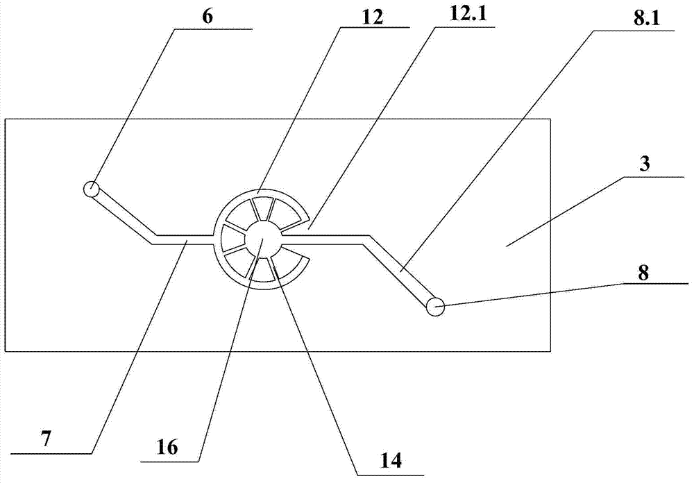
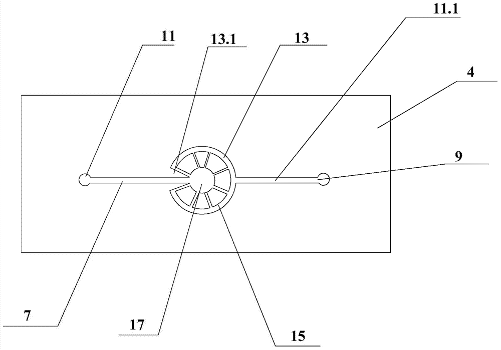
[0060] Such as Figure 1-6 Shown is a basic embodiment of a rare cell detection chip of the present invention. It includes a substrate 1 and a substrate 2 that is planarly consistent with the substrate 1 and sealed and bonded. The substrate 1 is composed of an upper layer 3, a lower layer 4 and a cell screening membrane 5 integrated between the upper layer 3 and the lower layer 4. One end of the upper layer 3 is provided with an upper layer sample inlet 6, and the other end is provided with an upper layer sample outlet 8. The upper layer sample inlet 6 is connected to an upper layer annular channel 12 with an upper layer gap 12.1 through the upper layer sample input channel 7, and the upper layer annular channel 12 passes through Several upper layer diffusion channels 14 that are radially arranged are connected to an upper layer circular sample pool 16, and this upper layer circular sample pool 16 is connected to the upper layer sample outlet 8 through the upper layer sample o...
Embodiment 2
[0066] Such as Figure 7 , 8 Shown is an embodiment of a rare cell detection chip of the present invention.
[0067] Figure 7 It is a scanning electron microscope image of the tapered micropore array filter membrane, wherein (a) is the side with the small diameter of the tapered micropores, and (b) is the side with the large diameter of the tapered micropores. The cell screening membrane 5 has a thickness of 40 micrometers. The thickness of the fixing ring 21 is 60 microns. The densely distributed and regularly arranged conical micropores on the cell screening membrane 5 have a small diameter of 6.5 microns, a large diameter of 25 microns, and a period of 40 microns. The material for making the microporous array filter membrane is polyethylene glycol.
[0068] Figure 8 for figure 1 The physical picture of the rare cell detection chip. The upper sample input channel 7, the upper sample output channel 8.1, the lower sample input channel 10, and the lower sample output ...
Embodiment 3
[0070] Using the rare cell detection chip in Example 2 to capture the colorectal cancer cell HT29 mixed in PBS solution includes the following steps:
[0071] 1) Sterilization: put the rare cell detection chip under ultraviolet light for sterilization;
[0072] 2) Surface modification of cell screening membrane 5: first inject 20 μl of MaxGel solution (diluted in PBS solution at a ratio of 1:100) into the chip with a syringe pump, incubate in the cell incubator for one hour, remove the residual solution, and then inject Phosphate buffered saline for washing;
[0073] 3) Cell capture: inject 10ml of phosphate buffer solution containing rectal cancer cell HT29 into the chip, so that the cancer cells are captured by the cell screening membrane 5;
[0074] 4) Cell fixation: inject a 4% paraformaldehyde solution into the chip to fix the captured cells on the cell screening membrane 5, and then inject phosphate buffer for washing;
PUM
| Property | Measurement | Unit |
|---|---|---|
| width | aaaaa | aaaaa |
| height | aaaaa | aaaaa |
| diameter | aaaaa | aaaaa |
Abstract
Description
Claims
Application Information
 - R&D
- Intellectual Property
- Life Sciences
- Materials
- Tech Scout
- Unparalleled Data Quality
- Higher Quality Content
- 60% Fewer Hallucinations
Browse by: Latest US Patents, China's latest patents, Technical Efficacy Thesaurus, Application Domain, Technology Topic, Popular Technical Reports.
© 2025 PatSnap. All rights reserved.Legal|Privacy policy|Modern Slavery Act Transparency Statement|Sitemap|About US| Contact US: help@patsnap.com


