Quick separation method of dendritic cells

A technology of dendritic cells and separation method, which is applied in the field of dendritic cell separation based on nano-magnetic beads, can solve the problems of spatial orientation change, cell extrusion, formation of precipitation, etc., to increase the chance of contact and realize cascade The effect of amplification, stabilization of the reaction solution

Inactive Publication Date: 2013-09-04
NANCHANG UNIV
View PDF1 Cites 1 Cited by
  • Summary
  • Abstract
  • Description
  • Claims
  • Application Information

AI Technical Summary

Problems solved by technology

However, the current separation technology based on micron-scale immunomagnetic beads has many limitations: 1) The specific surface area of ​​micron-sized magnetic beads is relatively small, which reduces the capture efficiency of magnetic beads; The combination of cells through a multiphase reaction usually takes longer to specifically capture the cells in the food matrix; 3) The micron magnetic beads have poor monodispersity and are prone to self-aggregation or self-aggregation in the whole blood matrix Precipitation; 4) The traditional immunomagnetic separation technology often directly couples the antibody to the immunomagnetic beads. This process often leads to a greatly reduced activity of the antibody and a change in the spatial direction of the antibody, which increases the inter-antibody interaction. Steric hindrance effect, which reduces the capture efficiency of antibodies 5) Blood viscosity is high and the concentration of non-DC blood cells is large, micron magnetic beads are prone to non-specific adsorption, and it is difficult to achieve specific separation of DC in blood; 6) Excessive concentration of micron magnetic beads will cause DC damage (the magnetic field causes the magnetic beads on the cell surface to attract each other, causing the cells to be squeezed or even ruptured), resulting in failure of separation
The distance between the antibody and the surface of the magnetic bead is too close, the nature of the magnetic bead itself and the residual hydrophobic or strong hydrophilic groups on the surface are likely to cause changes in the spatial conformation of the antibody, resulting in a decrease in the biological activity of the antibody

Method used


Image

  • Quick separation method of dendritic cells
  • Quick separation method of dendritic cells

Examples


Embodiment 1

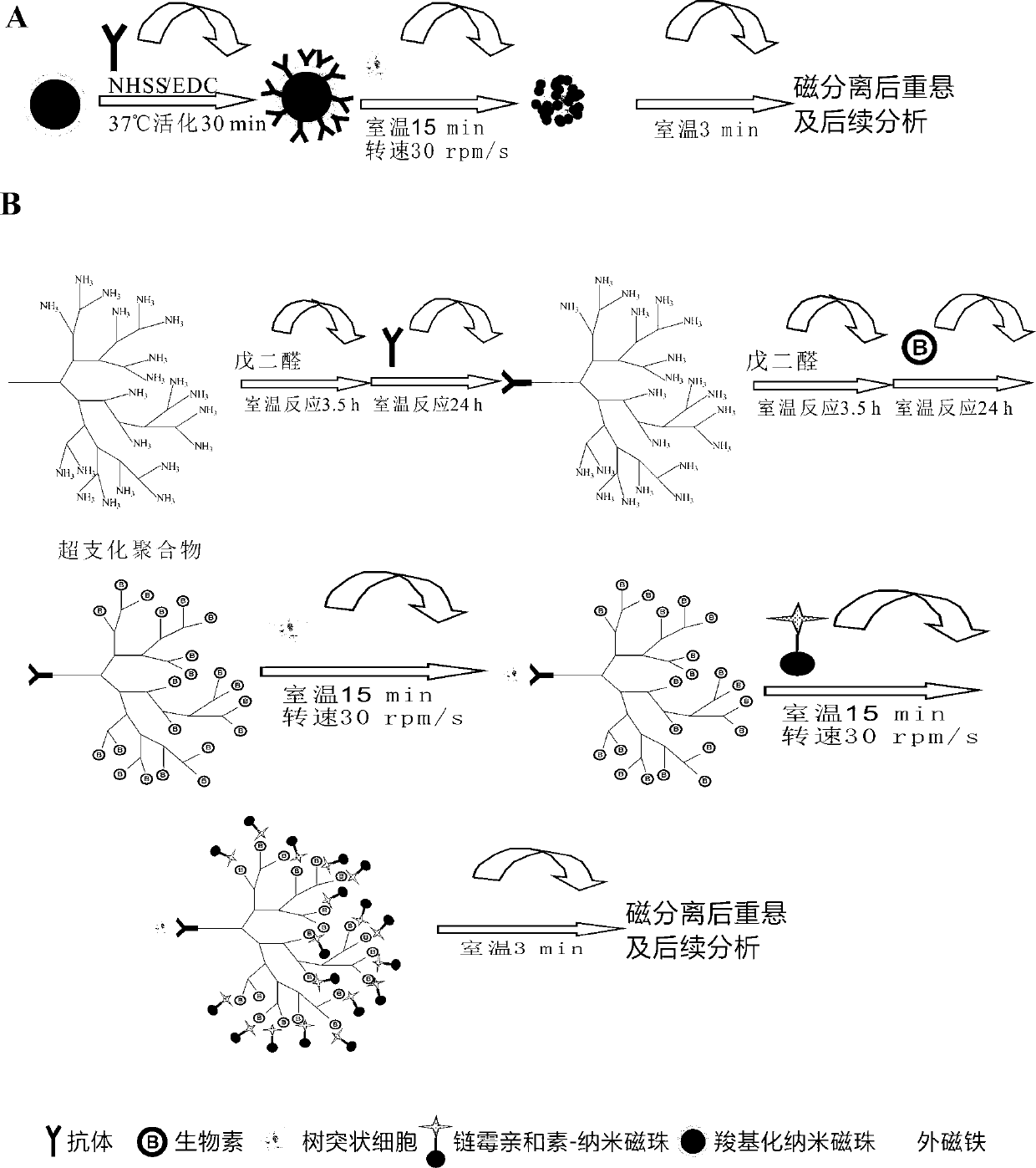
[0029] 1. The hyperbranched polymer-antibody complex is prepared according to the following steps:

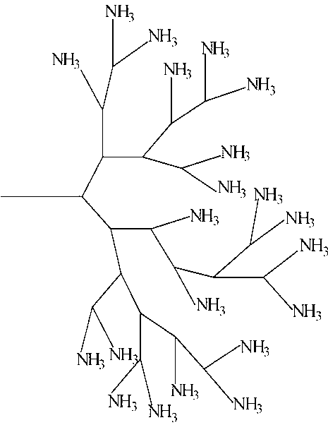
[0030] (1) Weigh 1.0 mg hyperbranched polyamidoamine aminated by hyperbranched polymer, suspend in 4 mL phosphate buffer (PBS, 0.01 mol / L, pH 8.0), stir and add 25% glutaraldehyde dropwise 545 μL of aqueous solution to make the final concentration of glutaraldehyde 3%. React at room temperature for 3.5 h at a rotating speed of 150 r / min on a shaker;

[0031] (2) Add 1 mL (18.75 mg) of mouse anti-human dendritic cell monoclonal antibody dropwise to the above solution so that the final concentration reaches about 3 mg / mL. React at room temperature for 24 h at the speed of the shaker at 150 r / min;

[0032] (3) The above solution was spin-dried under reduced pressure, dissolved in deionized water, and dialyzed in PBS and deionized water for 1 day; after the dialysis, the obtained solution was freeze-dried.

[0033] 2. The long-chain biotin-hyperbranched polymer-antibody...

Embodiment 2

[0039] Example 2 Enrichment effect experiment

[0040] (1) Take 1 mL of concentration as 10 4 Cells / mL DC cells were placed in a 1.5 mL sterile centrifuge tube, centrifuged at 12,000 rpm for 5 min, discarded the supernatant, and resuspended with an equal volume of sterile PBS solution.

[0041] (2) Enrichment and capture: Set up the technical scheme group of the present invention (hyperbranched polymer group co-modified with DC cell antibody and long-chain biotin), nano magnetic beads group modified with DC cell-specific antibody, and DC cell-specific antibody group Modified micron magnetic bead sets enrich target cells.

[0042] (3) After magnetic separation, pour the supernatant into a sterile centrifuge tube, and wash the isolated immunomagnetic beads with DC cells twice with PBST, mix well, and resuspend the immunomagnetic beads with 1 mL of sterile PBS solution. Magnetic bead complex.

[0043] (4) Capture rate calculation: after serial dilution of the enriched target ...

Embodiment 3

[0056] Example 3 Enrichment capture experiment

[0057] Conventional magnetic stand separation time is 30min, and all the other are with embodiment 2.

[0058] The catch rate of each group is as follows:

[0059] Capture rate of DC cell-specific antibody modified micron magnetic bead set Capture rate of DC cell-specific antibody-modified nanomagnetic bead set Capture efficiency of DC cell antibody and long-chain biotin co-modified hyperbranched polymer group 54.6% 40.1% 91.8%

[0060] The experimental results show that compared with the separation of 3 minutes in Example 2, when the separation time reaches 30 minutes, the capture efficiency of the three groups has been improved, especially the capture efficiency of the DC cell-specific antibody-modified nano-magnetic bead group is the most obvious, which shows that It shows that the capture efficiency of the nano-magnetic bead group can be greatly improved by extending the time, but it is still lower t...


PUM

PropertyMeasurementUnit
particle diameteraaaaaaaaaa

Abstract

The invention discloses a method for separating and enriching dendritic cells (Dendritice cells, DC) in whole blood for further providing a basis for subsequent study of dendritic cells, and relates to the biomedical field. The method comprises the following steps of: covalent coupling of hyperbranched polymers and mouse anti-human dendritic cell monoclonal antibodies; further coating long-chain biotin molecules by hyperbranched polymers modified by mouse anti-human dendritic cell monoclonal antibodies; capturing DC in a whole blood sample by hyperbranched polymers co-modified by mouse anti-human dendritic cell monoclonal antibodies and long-chain biotin; identifying and coupling long-chain biotin hyperbranched polymers in the whole blood by streptavidin modified nanomagnetic beads; and separating and resuspending the captured DC. The resuspension can be directly used for subsequent analysis. Compared with conventional cell separation methods, the method is more suitable for magnetic separation of DC in complex whole blood samples, so that the magnetic separation time is shortened, and the DC separation efficiency in the whole blood sample is improved.

Description

technical field [0001] The invention relates to the field of biomedicine, in particular to a method for separating dendritic cells based on nano magnetic beads. Background technique [0002] Dendritic cells (dendritic cells, DCs) were discovered by American scholar Steinman in 1973. They are the most powerful and only professional antigen presenting cells (antigen presenting cells, APCs) that can activate naive T cells. , its quantity and mature state can determine the immune status of the body, so its in-depth study has important theoretical and practical significance. DC is operable in vitro, so obtaining a certain amount of functional DC is the key to in-depth study of its biological characteristics. However, the content of DC in tissues is very small, accounting for only 0.16-0.68% of peripheral blood leukocytes and 0.5-1.6% of monocytes in blood. The existing methods cannot directly detect trace amounts of DC in peripheral blood. Therefore, the high-efficiency enrichm...

Claims


Application Information

Patent Timeline
Patent Type & Authority Applications(China)
IPC IPC(8): C12N5/0784
Inventor 许恒毅
Owner NANCHANG UNIV
Why Eureka
Try Eureka