Method for analyzing velocity vector of flow field of heart based on gray scale ultrasound image

A gray-scale ultrasound and velocity vector technology, applied in the field of medical image processing and cardiac fluid mechanics research, can solve the problems of slow blood flow, no multi-scale weight optimization, and inability to obtain motion vector results, etc., to achieve smooth blood flow area Effect

Active Publication Date: 2013-02-13
SICHUAN PROVINCIAL PEOPLES HOSPITAL
View PDF3 Cites 8 Cited by
  • Summary
  • Abstract
  • Description
  • Claims
  • Application Information

AI Technical Summary

Problems solved by technology

[0008] VFM method: VFM method is a method applied to Doppler color Doppler ultrasound to calculate the motion vector. Due to the inherent angle dependence of the algorithm, the clinical application is very unstable, and it cannot process gray-scale ultrasound images.
[0009] Optical flow method: It is extremely rare to apply optical flow method to the study of black and white ultrasound images of the heart. P. Burlina et al. applied the existing optical flow method to ultrasound contrast images in 2011, but the effect was not very satisfactory. On the one hand, it did not tar

Method used


Image

  • Method for analyzing velocity vector of flow field of heart based on gray scale ultrasound image
  • Method for analyzing velocity vector of flow field of heart based on gray scale ultrasound image
  • Method for analyzing velocity vector of flow field of heart based on gray scale ultrasound image

Examples


Embodiment

[0043] See attached figure 1 , the cardiac flow field velocity vector analysis method based on the gray-scale ultrasonic image provided by the present invention comprises the following steps:

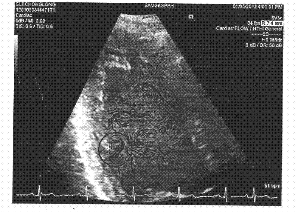
[0044] ①The grayscale ultrasound image with a size of 520×512 (such as figure 2 and image 3 As shown), the horizontal and vertical divisions are equally spaced into 7×7 grid units; wherein, the horizontal and vertical spacing can be the same or different, and here, the horizontal and vertical division spacings are both 7 pixels;

[0045] ② Calculate the average gray value P of each cell in the grid unit mn, and select the pixel point closest to the average gray value in the cell as the control point of the cell; where the calculation formula of the average gray value is:

[0046] P mn = Σ i , j ...


PUM


Abstract

The invention discloses a method for analyzing the velocity vector of a flow field of a heart based on a gray scale ultrasound image. The method is characterized by comprising the following steps of: dividing the gray scale ultrasound image into a grid unit; selecting a pixel point of which the pixel is closest to the average scale value in each cell of the grid unit, wherein the pixel point is used as a control point, and updating the gray value of the gray scale ultrasound image by an interpolation algorithm to fulfill the aim of removing speckle noise; and calculating by an optical flow method to obtain the heart flow field movement vector of the denoised gray ultrasound image. According to the method, multiple times of recursive computation is not needed in the interpolation algorithm process, and speckles can be removed and preprocessed in real time; and a calculation result is closest to the actual condition, and detailed information of flow such as eddy can be accurately acquired.

Description

technical field [0001] The invention relates to the fields of medical image processing and heart fluid mechanics research, in particular to a heart flow field velocity vector analysis method based on gray-scale ultrasonic images. Background technique [0002] Synchronously extracting the dynamic motion vectors of blood and myocardium from cardiac ultrasound image sequences involves two important steps: removing the influence of speckle noise on the image and calculating motion vectors; [0003] (1) Ultrasound image denoising: The purpose of medical noise image denoising is to maintain the important features of the original image as much as possible while smoothing the noise. The noise of medical ultrasound images is mainly speckle noise, and the academic community has proposed some methods to suppress speckle noise to provide high-quality images for subsequent processing. Spatial filtering of speckle noise includes mean filtering, median filtering, Lee filtering, Kuan filte...

Claims


Application Information

Patent Timeline
IPC IPC(8): G06T5/00G06T7/00
Inventor 甘建红尹立雪谢盛华
Owner SICHUAN PROVINCIAL PEOPLES HOSPITAL
Why Eureka
Try Eureka