Near-infrared fluorescent nanoparticle in-vivo probe and preparation method thereof

A near-infrared and internal fluorescence technology, applied in the field of preparation of near-infrared in vivo fluorescent nanoparticle probes and near-infrared in vivo fluorescent nanoparticle probes, can solve the problem of reducing the fluorescence intensity of quantum dots in vivo, harsh reaction conditions, inapplicability And other issues

Inactive Publication Date: 2013-01-16
CHINA PHARM UNIV
View PDF2 Cites 8 Cited by
  • Summary
  • Abstract
  • Description
  • Claims
  • Application Information

AI Technical Summary

Problems solved by technology

However, at present, there are still some problems to be overcome in terms of biocompatibility, stability, and toxicity of ordinary near-infrared quantum dots alone.
[0004] In order to improve the stability and toxicity of ordinary quantum dots, the usual method is to modify the surface of quantum dots, prepare functionalized quantum dots or pack them into polymer microspheres, but these methods often reduce the Fluorescence intensity or in vivo stability, some polymer microspheres are not suitable for in vivo imaging due to their large size, and they also need to use some expensive reagents or very harsh reaction conditions

Method used


Image

  • Near-infrared fluorescent nanoparticle in-vivo probe and preparation method thereof
  • Near-infrared fluorescent nanoparticle in-vivo probe and preparation method thereof
  • Near-infrared fluorescent nanoparticle in-vivo probe and preparation method thereof

Examples


Embodiment 1

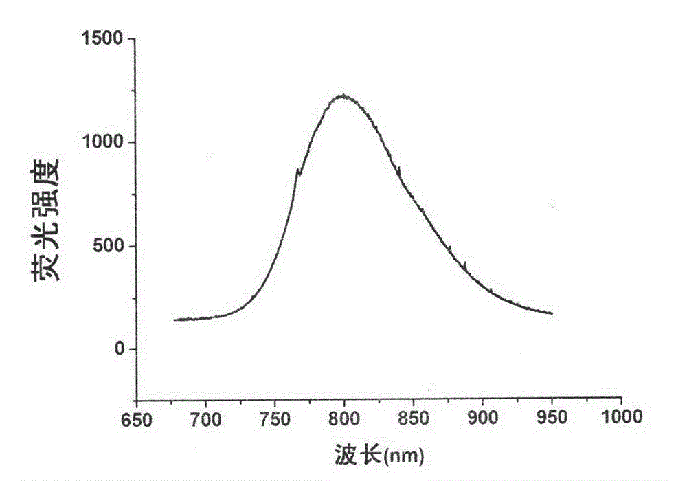
[0033] Step 1: Preparation of water-soluble near-infrared CdHgTe fluorescent quantum dots

[0034] Accurately weigh 60mg of tellurium powder, NaBH 4 40mg, placed in a 25ml three-necked bottle, under N 2 Under air protection, 2 ml of double distilled water was added, and reacted in a water bath at 40° C. for 30 minutes to obtain a purple-red NaHTe solution. N was introduced into the solution 2 The solution was deoxygenated by air for 30 min. Another preparation 100ml containing 92.4mgCd(NO 3 ) 2 solution, and added 63 μl of mercaptoacetic acid (MPA) as a stabilizer, adjusted the pH to 11.2 with 1mol / L NaOH, and added 10 μl of mercuric nitrate solution. N was introduced into the solution 2 The solution was deoxygenated by air for 30 min. Under vigorous stirring, inject 600 μl of the above-mentioned deoxygenated 0.5mol / L NaHTe solution to make Cd 2+ : NaHTe: The molar ratio of MPA is 1: 0.5: 2.4, then rapidly heated to reflux to boiling. Reflux for 1 hour to obtain a CdH...

Embodiment 2

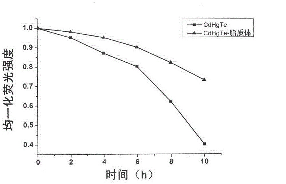
[0040] (1) Stability investigation of CdHgTe quantum dots and CdHgTe quantum dots-liposome fluorescent probes:

[0041]The CdHgTe quantum dots prepared in Example 1 and the CdHgTe quantum dot-liposome fluorescent probes were all placed under 20W254nm ultraviolet lamps for irradiation, and the fluorescence intensity was measured at 0, 2, 4, 6, 8, and 10 hours, and compared the two resistance to photobleaching. see results image 3 . It can be seen that the photostability of the CdHgTe quantum dot-liposome fluorescent probe is obviously better than that of the single CdHgTe quantum dot.

[0042] (2) Toxicity investigation of CdHgTe quantum dots and CdHgTe quantum dots-liposome fluorescent probes:

[0043] Select human breast cancer cell MCF-7 cells, add them into 96-well culture plate, set up five duplicate wells, set at 37°C, 5% CO 2 Cultivate overnight in the incubator, replace the culture medium with 0.5% serum M199 culture medium to synchronize the cells, then add the Cd...


PUM


Abstract

The invention relates to a near-infrared fluorescent nanoparticle in-vivo probe. The near-infrared fluorescent nanoparticle in-vivo probe comprises water-soluble near-infrared CdHgTe fluorescent quantum dots and liposomal phospholipid film wrapped outside the dots. The invention further provides a preparation method of the near-infrared fluorescent nanoparticle in-vivo probe. The preparation method includes the steps of firstly, synthetizing the water-soluble near-infrared CdHgTe fluorescent quantum dots; secondly, preparing the liposomal phospholipid film; and thirdly, adding water-soluble near-infrared fluorescent quantum dots CdHgTe solution synthetized in the step 1 into the liposomal phospholipid film prepared in the step 2 to obtain the near-infrared fluorescent nanoparticle in-vivo probe. The near-infrared fluorescent nanoparticle in-vivo probe is high in stability and biocompatibility. The preparation process is simple, the cost is low, and the near-infrared fluorescent nanoparticle in-vivo probe is convenient to use and effective in in-vivo imaging.

Description

technical field [0001] The invention relates to the field of near-infrared living body imaging, in particular to a near-infrared in vivo fluorescent nanoparticle probe, and at the same time, the invention also provides a preparation method for the near-infrared in vivo fluorescent nanoparticle probe. Background technique [0002] Near-infrared imaging technology is a new spectral imaging method developed in recent years. In the range of near-infrared band (700-900nm) applied by this technology, it avoids the main absorption area of ​​many endogenous substances such as oxygenated hemoglobin, anoxygenated hemoglobin, water, etc., so the penetration depth in biological tissues is relatively large (up to 10cm), and the biological autofluorescence in this band is also reduced to the lowest level, and the imaging signal is less affected by the background of biological tissues. The potential of near-infrared imaging technology for in-situ non-destructive and continuous monitoring ...

Claims


Application Information

Patent Timeline
IPC IPC(8): C09K11/89C09K11/02A61K49/00
Inventor 胡育筑叶超李臣贵
Owner CHINA PHARM UNIV
Why Eureka
Try Eureka