Method for detecting microcystin in water
A technology of microcystin and detection method, which is applied in measurement devices, instruments, Raman scattering, etc., can solve the problems of expensive equipment, large sample consumption, inability to identify MC species, etc., and achieve the effect of sensitive detection
- Summary
- Abstract
- Description
- Claims
- Application Information
AI Technical Summary
Problems solved by technology
Method used
Image
Examples
preparation example Construction
[0025] (1) Preparation of MAb-MPs solution
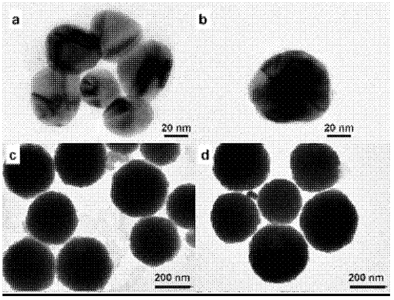
[0026] Firstly prepare magnetic nanoparticles (select ferric oxide nanoparticles in this embodiment), prepare by hydrothermal method, the specific steps are as follows: 0.45g~0.75g ferric chloride, 0.10g~0.20g trisodium citrate are dissolved in 20ml ~40ml of ethylene glycol, then add 1.0g~2.0g of sodium acetate, stir for 30min, pour the resulting mixture into a 50ml reaction kettle, and react at 200°C for 10h; Wash with ethanol three times, and finally dissolve in water to obtain an aqueous solution of iron ferric oxide nanoparticles with a diameter of 180nm to 250nm. figure 1 as shown in (c);
[0027] Microcystin monoclonal antibodies (MAbs), respectively MC-LR, MC-YR and MC-RR, were linked to the prepared magnetic nanoparticles with EDC-NHS coupling agent, the specific steps are as follows : Mix 20 μL of a molar concentration of 3.0 mM NHS (N-hydroxysuccinimide) and 20 μL of a molar concentration of 3.0 mM EDC (1-(3-dimethylamin...
Embodiment 1
[0037] Embodiment 1, the detection of microcystin MC-LR in water
[0038] (1) Drawing of detection standard curve
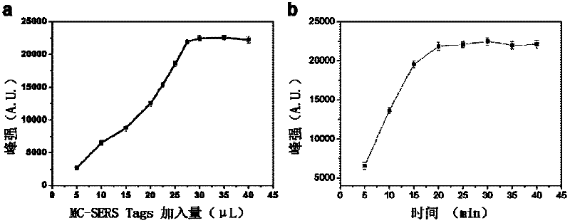
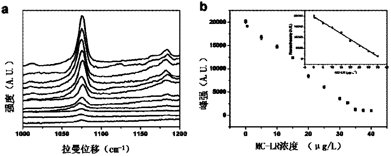
[0039] 10 μL of MC-LR standard solutions of different concentrations (concentrations are 0 μg / L, 0.02 μg / L, 0.04 μg / L, 0.06 μg / L, 0.1 μg / L, 0.5 μg / L, 1 μg / L, 1.5 μg / L L, 2μg / L, 2.5μg / L, 3μg / L, 3.5μg / L and 4μg / L) were mixed with 27.5μL of MC-SERS Tags (connected with MC-LR molecule) solution in turn to obtain the incubation solution; solution was added to 20 μL of MAb-MPs (MC-LR monoclonal antibody) solution and shaken for 25 minutes, the magnetic immune complex in the solution was separated with a magnet, the remaining solution was discarded, and the obtained immune complex was washed with PBS Wash twice, then detect its Raman signal with a laser Raman spectrometer, (laser Raman detects the incident light wavelength is 532nm, the power is 2mW, and the spectral detection time is 10s); when using different MC-LR concentrations, the Raman spectrum is 1076cm -1 The...
Embodiment 2
[0042] Embodiment 2, the detection of microcystin MC-YR in water
[0043] (1) Drawing of detection standard curve
[0044] 10 μL of different concentrations of MC-YR standard solution (concentrations are 0 μg / L, 0.02 μg / L, 0.04 μg / L, 0.06 μg / L, 0.1 μg / L, 0.5 μg / L, 1 μg / L, 1.5 μg / L L, 2μg / L, 2.5μg / L, 3μg / L, 3.5μg / L and 4μg / L) were mixed with 27.5μL of MC-SERS Tags (connected with MC-YR molecule) solution in turn to obtain the incubation solution; solution was added to 20 μL MAb-MPs (connected with MC-YR monoclonal antibody) solution and reacted for 25 minutes by shaking, the magnetic immune complex in the solution was separated with a magnet, the remaining solution was discarded, and the obtained immune complex was washed with PBS Wash twice, then detect its Raman signal with a laser Raman spectrometer, (laser Raman detects the incident light wavelength is 532nm, the power is 2mW, and the spectral detection time is 10s); when using different MC-YR concentrations, the Raman spe...
PUM
| Property | Measurement | Unit |
|---|---|---|
| Particle size | aaaaa | aaaaa |
| Particle size | aaaaa | aaaaa |
| Thickness | aaaaa | aaaaa |
Abstract
Description
Claims
Application Information
 - R&D Engineer
- R&D Manager
- IP Professional
- Industry Leading Data Capabilities
- Powerful AI technology
- Patent DNA Extraction
Browse by: Latest US Patents, China's latest patents, Technical Efficacy Thesaurus, Application Domain, Technology Topic, Popular Technical Reports.
© 2024 PatSnap. All rights reserved.Legal|Privacy policy|Modern Slavery Act Transparency Statement|Sitemap|About US| Contact US: help@patsnap.com


