Activation and monitoring of cellular transmembrane potentials
一种跨膜电位、膜电位的技术,应用在监测和操纵细胞跨膜电压的组合物,纳米颗粒和它们在监测和操纵跨膜电压中的应用领域,能够解决低假阴性假阳性”率等问题,达到高量子产率的效果
- Summary
- Abstract
- Description
- Claims
- Application Information
AI Technical Summary
Problems solved by technology
Method used
Image
Examples
Embodiment 1
[0129] Labeling membranes with quantum dots
[0130] Quantum dots are ideal candidates for use as voltage-sensitive probes because their physical size is comparable to the thickness of cell membranes and their electronic properties make them potentially tunable by external electromagnetic fields. refer to Figure 6A , the absorption of a photon 602 having a first wavelength and having a higher energy than the band gap 604 between the HOMO valence band 606 and the LUMO conduction band 608 of the quantum dot forms electron-hole pairs, which can radiatively recombine to emit photons 610 having the second wavelength. Strong local electric fields (eg, due to changes in membrane potential across cellular lipid bilayers) can interact with free charge carriers generated by excited quantum dots. Figure 6B It is schematically illustrated how the electric field 612 can adjust the optical properties of the emitted light 614 . For example, a membrane potential of 100 mV across a hydrop...
Embodiment 2
[0133] Screening test for quantum dots
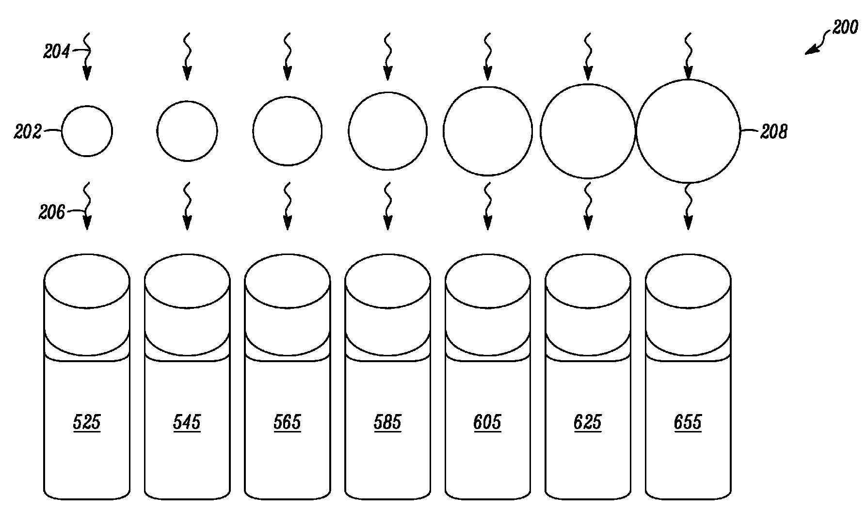
[0134] A QDOT nanocrystal-based optical voltage sensor is provided to monitor rapid changes in cell membrane potential. When located within the cell membrane, the nanoparticles are sensitive to and detect changes in the transmembrane voltage gradient and report it as a change in their emission properties. A series of quantum dots were prepared and screened to monitor their optical properties in response to chemical and electrical stimuli of cells.
[0135] A. Effects of chemical stimuli
[0136] The effects of chemical stimuli on quantum dot-treated cells were monitored according to the protocol described below. Cells were incubated with the nanocrystal-based voltage sensor for 1 hour in a 96-well plate, after which any excess nanoparticles were washed away. Depolarize the labeled cell population using a chemical stimulus (100 mM KCl) suitable for high-volume imaging experiments. Figure 10 The emission intensity (expressed as rel...
Embodiment 3
[0141] Quantum dot-based activation platform
[0142]A light-controlled activation platform for long-range and reversible manipulation of membrane potential is described. The materials and methods described provide an alternative to passively monitoring the functional activity of cells and provide the ability to non-invasively manipulate the membrane potential of cells, which is critical for understanding their development, communication and fate. Traditional non-physiological approaches most commonly rely on pharmacological interventions (by direct stimulation of cells with the addition of "high K+" solutions or pharmacological channel openers or with microelectrodes). However, these non-physiological approaches have serious drawbacks, including lack of control of the membrane potential, irreversibility of the induced changes, and low temporal resolution.
[0143] The alternative presented here uses light to trigger long-range reversible manipulation of membrane potential in...
PUM
 Abstract
Description
Claims
Application Information
 - R&D
- Intellectual Property
- Life Sciences
- Materials
- Tech Scout
- Unparalleled Data Quality
- Higher Quality Content
- 60% Fewer Hallucinations
Browse by: Latest US Patents, China's latest patents, Technical Efficacy Thesaurus, Application Domain, Technology Topic, Popular Technical Reports.
© 2025 PatSnap. All rights reserved.Legal|Privacy policy|Modern Slavery Act Transparency Statement|Sitemap|About US| Contact US: help@patsnap.com


