Fluorescent microsphere immunochromatography detection card for detecting enrofloxacin and preparation method thereof
A technology of fluorescent microspheres and enrofloxacin, which is applied in chemical instruments and methods, measuring devices, and analytical materials, etc., can solve problems such as inability to sample multiple dilutions, sensitivity limitations, and many interfering components, and achieve low prices, The effect of high sensitivity and increased fluorescence lifetime
- Summary
- Abstract
- Description
- Claims
- Application Information
AI Technical Summary
Problems solved by technology
Method used
Image
Examples
Embodiment 1
[0043] 1. Preparation of fluorescent microsphere markers (EDC method):
[0044] Preparation of fluorescent microsphere-labeled anti-enrofloxacin antibody: Take 1 mg of fluorescent microspheres coated with isothiocyanatorhodamine organic dye and centrifuge at 1000×g for 10 minutes, collect the precipitate, and wash with 0.01M borate buffer solution of pH 4.8 Adjust the microsphere concentration to OD 450 =0.2, then add 90μl 50mg / ml EDC, 150μl 5mg / ml NHS, shake and mix well, incubate at room temperature for 20min, centrifuge at 1000×g for 5min, dissolve the precipitate with 0.01M borate buffer solution of pH4.8, and adjust micro Ball concentration is OD 450 is 0.5. at 0.1ml OD 450 Add 1 μg of anti-enrofloxacin antibody to the fluorescent microspheres with a pH of 0.5, mix well, stir and react at room temperature for 3 h, wash and centrifuge 3 times with ultrapure water, wash with 0.01M PBS of pH 7.2 (which contains 5% sucrose and 0.05% Tween-20) to resuspend the pellet to th...
Embodiment 2
[0056] The preparation method of the present embodiment is basically the same as that of Example 1, the difference is:
[0057] The carrier protein is ovalbumin, and the labeled carrier fluorescent microspheres are quantum dot-silica core-shell dual-structure microspheres.
[0058] The qualitative detection of enrofloxacin with the above-mentioned fluorescent microsphere immunochromatographic detection card includes the following steps:
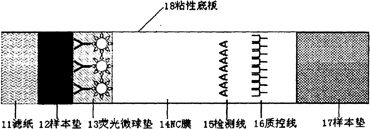
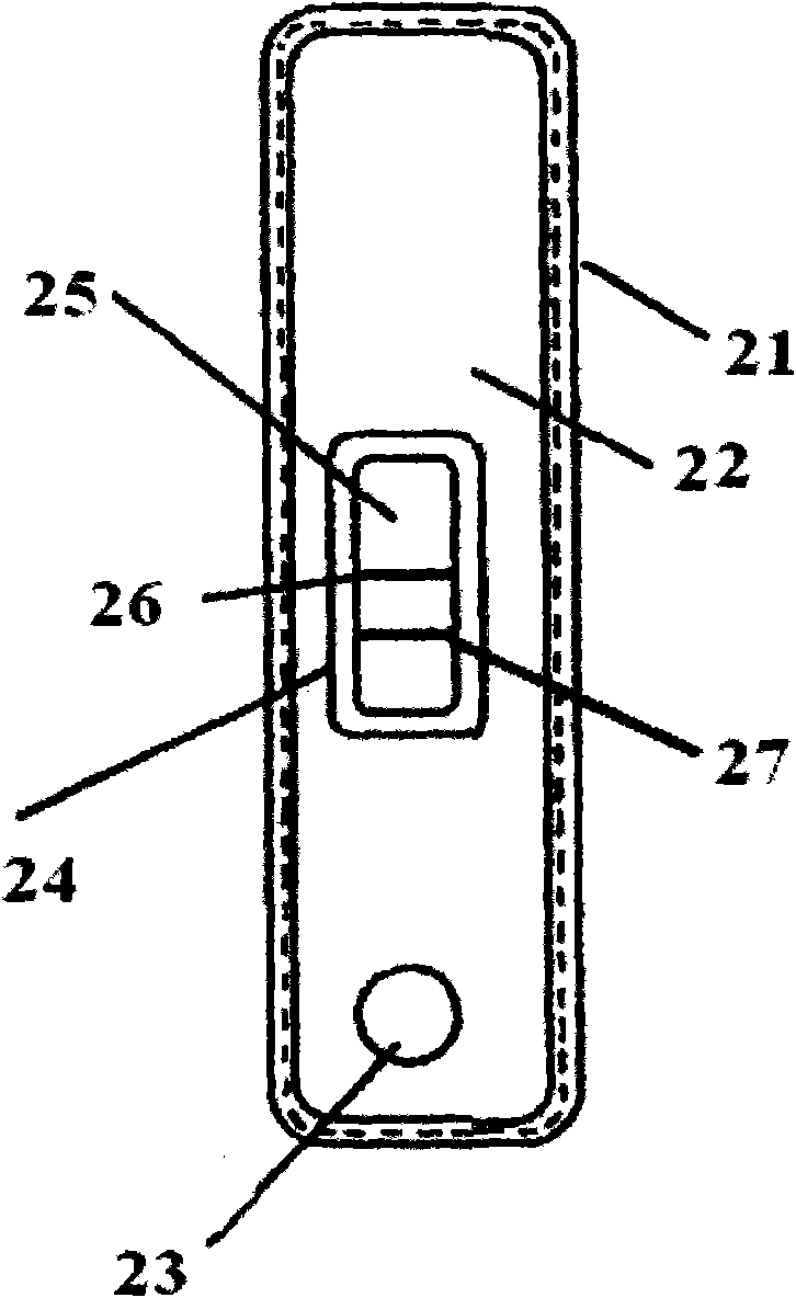
[0059] (1) Lay the test card flat, after the chicken liver extract sample to be tested is equilibrated to room temperature, add 90 μl of sample into the sample hole of the test card, react at room temperature for 15 minutes, and put the test card into the detection window;
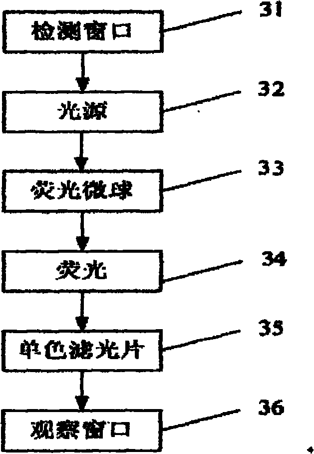
[0060] (2) The fluorescent microspheres trapped in the detection area and the quality control area emit strong fluorescence under the excitation of the best excitation light source; observe in the observation window, if there is a fluorescent band in the detection area of...
Embodiment 3
[0062] The preparation method of this example is basically the same as that of Example 1, the difference is: 1. The fluorescent microspheres of the labeling carrier are isothiocyanatofluorescein-silicon dioxide core double-structured microspheres, and the surface is modified by streptavidin and prime. 2. The anti-enrofloxacin antibody can be coupled with biotin by the EDC method, which is basically the same as in Example 1.
PUM
| Property | Measurement | Unit |
|---|---|---|
| diameter | aaaaa | aaaaa |
Abstract
Description
Claims
Application Information
 - R&D
- Intellectual Property
- Life Sciences
- Materials
- Tech Scout
- Unparalleled Data Quality
- Higher Quality Content
- 60% Fewer Hallucinations
Browse by: Latest US Patents, China's latest patents, Technical Efficacy Thesaurus, Application Domain, Technology Topic, Popular Technical Reports.
© 2025 PatSnap. All rights reserved.Legal|Privacy policy|Modern Slavery Act Transparency Statement|Sitemap|About US| Contact US: help@patsnap.com


