Medical application of 6-shogaol for treating cervical cancer, leukemia and breast cancer
A technology of shogaol and cervical cancer, which is applied in the direction of medical preparations containing active ingredients, pharmaceutical formulas, antineoplastic drugs, etc., and can solve the problems of weak activity and weak activity
- Summary
- Abstract
- Description
- Claims
- Application Information
AI Technical Summary
Problems solved by technology
Method used
Image
Examples
Embodiment 1
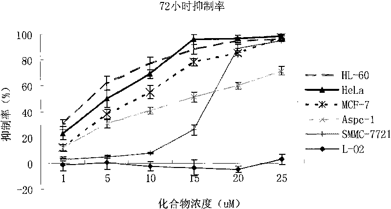
[0012] Antitumor effect of 6-shogaol in vitro
[0013] Take the cells in the logarithmic growth phase and inoculate 4~10×10 cells according to the size of the cells 3 Each was placed on a 96-well plate, and after 24 hours of growth, the supernatant was discarded, and then administered according to the following groups: Tumor cells were set up with no drug group and drug-added group (concentration 1-25 μM), and each group was set up with 5 or 6 replicates. Incubate wells for 24 or 72 hours, discard the supernatant, add 100 μl of MTT (tetrazolium salt) serum-free medium containing 0.5 mg / ml and incubate for 4 hours, add 100 μl DMSO (dimethyl sulfoxide), and place in a micro shaker Shake it for 10 minutes, and then place it on a microplate reader to detect the OD value at 570nm. Normal human cell line L-O2 was used for toxicity evaluation, and each experiment was repeated 3 times. The results are shown in Table 1 and Figure 1 ~ Figure 2 .
[0014] Table 1 IC of 6-shogaol on ...
Embodiment 2
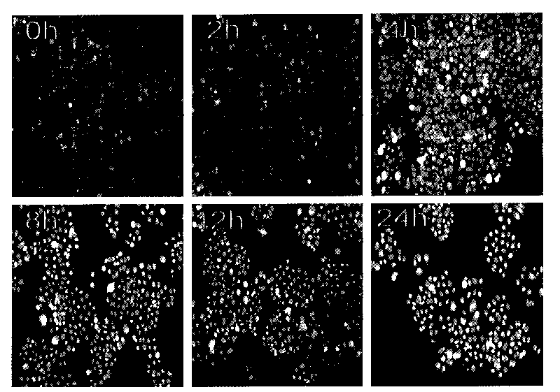
[0020] Cell morphology observation and AO (acridine orange) staining
[0021] Tumor cells were divided into no-drug group and drug-added group (5μM). Cells were treated for different time (0, 2, 4, 8, 12, 24 hours), fixed with 4% paraformaldehyde for 30 minutes, washed 2-3 times with 0.01M PBS, stained with AO for 10 minutes, and observed the morphology of the nucleus under a fluorescent microscope .
[0022] It was found that the morphology of the cells in the negative treatment group was normal, and the nuclei were intact after staining with the staining reagent. Many cells were rounded or shrunk after being treated with 6-shogaol, and the nuclei were found to be pyknotic when excited by blue light under a fluorescent microscope.
[0023] After AO fluorescent staining, the nuclei of typical apoptotic cells were densely stained and showed blocky or granular yellow-green fluorescence; normal cells were evenly stained; dead cells were not stained. The experimental results sho...
Embodiment 3
[0025] Detection of cell apoptosis by flow cytometry
[0026] In the experiment, a negative control group (ie no drug treatment group) and a 6-shogaol treatment group (15 μM) were set up. After the cells were treated with 6-shogaol for 12 hours, they were digested with 0.1% trypsin without EDTA. After the digestion was terminated by the serum-containing medium, the cells were collected in a 10ml glass centrifuge tube, centrifuged at 500rpm (or 100g) for 5min, discarded After washing with 0.01M PBS at 500rpm (or 100g) for 5min, centrifuge twice, then add the binding liquid according to the instructions of the Annexin V / PI double staining kit and perform staining for flow cytometry (Becton Dickinson FACScan Flow Cytometer) detection. FACS data were analyzed by professional flow cytometer operators using FLOWJO software (Tree Star, CA). The results showed that 6-shogaol could induce apoptosis.
[0027] The 18-hour apoptosis rate of the negative group without drugs was 4.8±1.7%....
PUM
 Abstract
Description
Claims
Application Information
 - R&D Engineer
- R&D Manager
- IP Professional
- Industry Leading Data Capabilities
- Powerful AI technology
- Patent DNA Extraction
Browse by: Latest US Patents, China's latest patents, Technical Efficacy Thesaurus, Application Domain, Technology Topic, Popular Technical Reports.
© 2024 PatSnap. All rights reserved.Legal|Privacy policy|Modern Slavery Act Transparency Statement|Sitemap|About US| Contact US: help@patsnap.com


