Method for producing light field or wide field fluorescence light section
A fluorescent light and slicing technology, applied in the field of optical slicing, can solve the problems of toxic damage to biological samples, affecting imaging speed, sample limitation, etc., and achieve the effects of wide application prospects, increased imaging speed, and simple settings.
- Summary
- Abstract
- Description
- Claims
- Application Information
AI Technical Summary
Problems solved by technology
Method used
Image

Examples
Embodiment 1
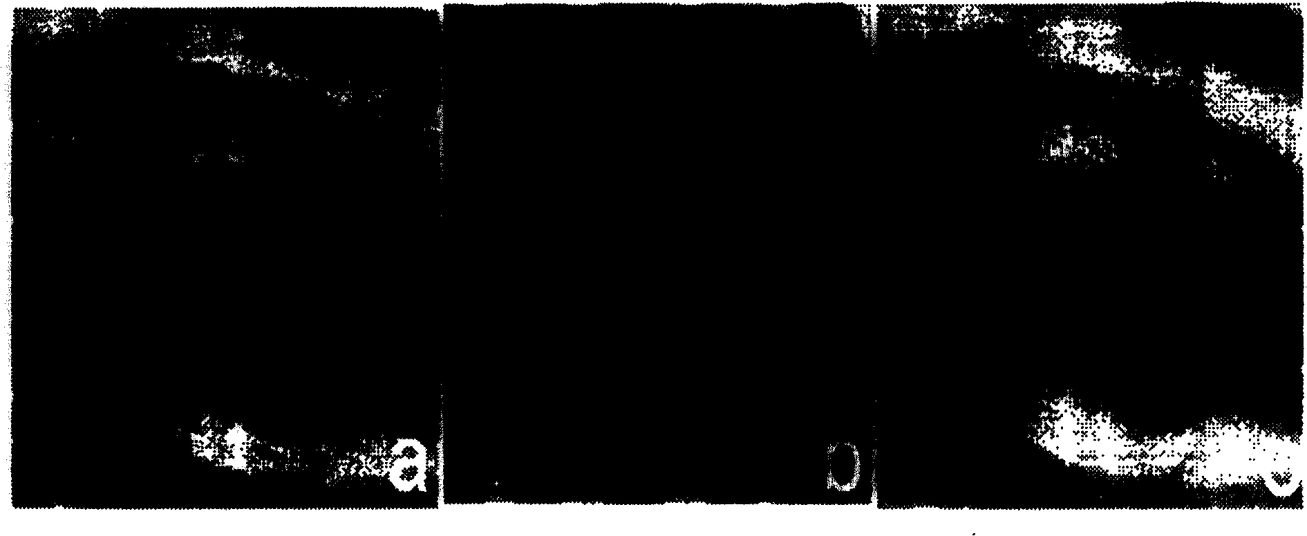
[0025] Observation of corneal slices and imaging of bright-field light slices are realized on an inverted microscope with the technology that can generate wide-field light slices. The objective lens used is a 40× plan achromatic objective lens. The specific method is: a 25 / mm sinusoidal grating is installed in front of the microscope tungsten halogen lamp (wide-field white light source), and the image square Fourier spectrum plane behind the microscope objective lens is Set a pass filter on it. The tungsten-halogen lamp projected the grating on the image of the corneal slice to obtain an optical slice image with a thickness of 0.7 microns. Adjust the focal length of the microscope objective lens to obtain the optical section images of each slice. A bandpass filter removes the grating fringes into a clear image of the light slice. Specific images such as figure 1 shown by figure 1 It can be seen that the resulting corneal slice images not only have clear boundaries, but als...
Embodiment 2
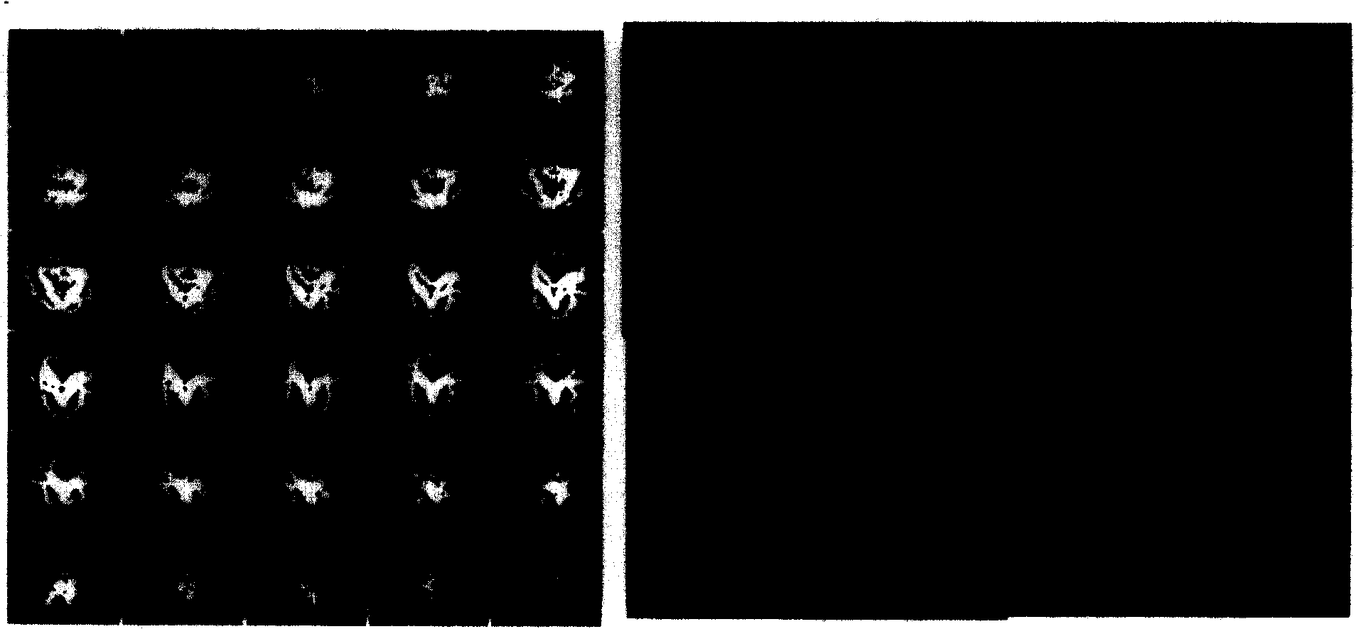
[0027] The tomographic wide-field optical slice imaging of pollen spores is realized on a fluorescence microscope by using the optical spatial filtering method that can generate three-dimensional images. The objective lens used is a 60× plan achromatic objective lens. The specific method is: set a 100 / mm sinusoidal grating in front of the microscope mercury lamp (wide-field ultraviolet light source). The mercury lamp projects the grating onto the sample for imaging, and the band-pass filter filters out the grating stripes to form a clear light slice image. Adjust the focal length of the microscope objective lens to obtain optical slice images of 30 slices, each with a thickness of 0.3 μm. Specific images such as figure 2 As shown, the images of each optical slice are clear, and the images of each layer transition gradually. figure 2 At the same time, the 3D stereoscopic image obtained after 3D reconstruction of the 30-layer optical slice image is given with our 3D image r...
Embodiment 3
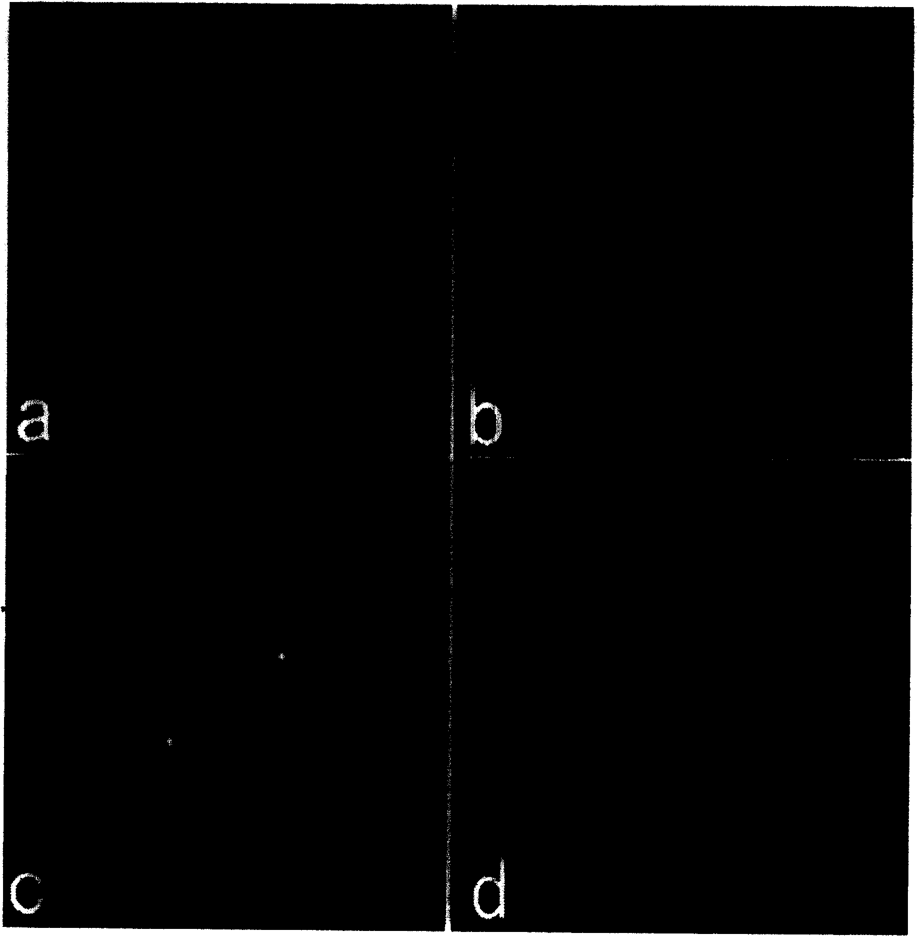
[0029] The tomographic wide-field optical slice imaging of fingerprints is realized on a polarizing microscope by means of an optical spatial filtering method that can generate three-dimensional images. The objective lens used is a 20× objective lens. The specific method is: set a 150 / mm sinusoidal grating in front of the microscope tungsten-halogen lamp (wide-field white light source), and the tungsten-halogen lamp projects the grating on the sample slide with fingerprints for imaging, and the resulting image is then processed by digital image to perform spatial filtering. Specific images such as image 3 as shown, image 3 a is an image superimposed with grating stripes, and the frequency spectrum of the image after Fourier transform after digital image processing is as follows: image 3 c see. image 3 d is to remove the part of the spectrum corresponding to the grating stripes by means of digital image processing, image 3 b is the tomographic optical slice image of t...
PUM
 Abstract
Description
Claims
Application Information
 - Generate Ideas
- Intellectual Property
- Life Sciences
- Materials
- Tech Scout
- Unparalleled Data Quality
- Higher Quality Content
- 60% Fewer Hallucinations
Browse by: Latest US Patents, China's latest patents, Technical Efficacy Thesaurus, Application Domain, Technology Topic, Popular Technical Reports.
© 2025 PatSnap. All rights reserved.Legal|Privacy policy|Modern Slavery Act Transparency Statement|Sitemap|About US| Contact US: help@patsnap.com