Automated microscopic cell analysis
- Summary
- Abstract
- Description
- Claims
- Application Information
AI Technical Summary
Benefits of technology
Problems solved by technology
Method used
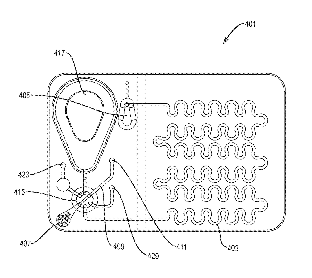
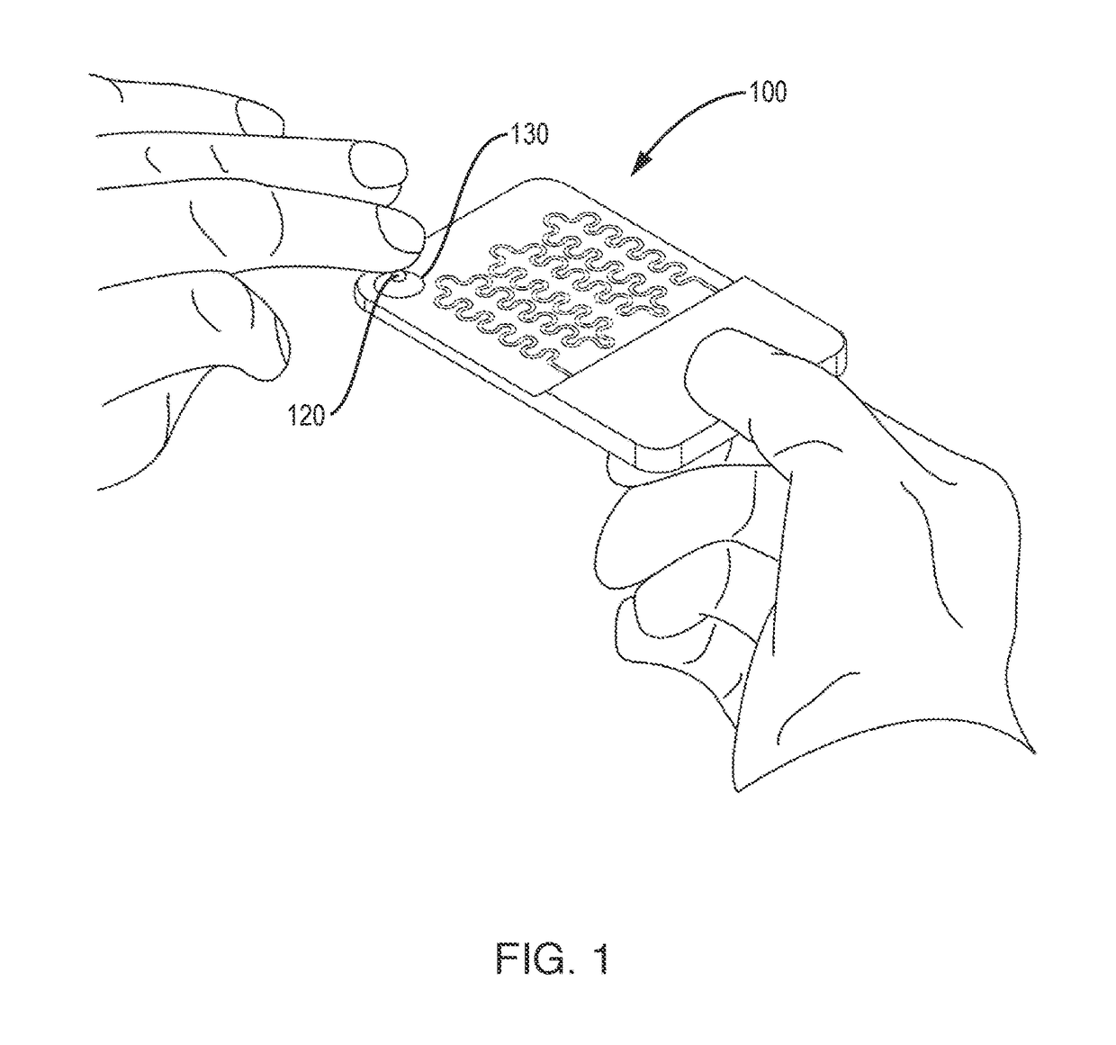
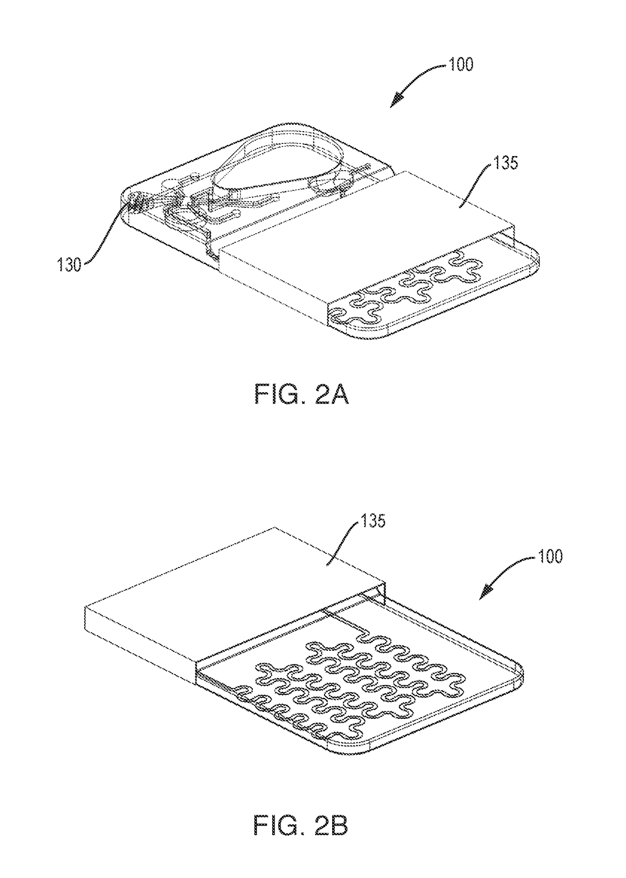
Image

Examples
example 1
on from Bright-Field and Fluorescent Optics
[0084]FIG. 12 shows images that were collected using test devices according to the present invention. A fluorescent stain Acridine Orange (AO) was used to differentially stain DNA and RNA of cells in a whole blood sample. The visual images of FIG. 12 were obtained using an Olympus 20××0.4 NA objective lens 265 and a Basler 5 MP digital camera 280. Excitation of the bright-field images in the second column was provided by white light bright-field source 260. Excitation of the fluorescent images in the third column was a 455 nm blue fluorescent light source 270.
[0085]White blood cells have significant RNA and DNA and therefore can be seen in the fluorescent images having green and orange structures. The size and shape of the green nuclear structure and overall size of the white cells can be used to differentiate them into sub-groups identified by name in the first column. Notably the basophil and eosinophil sub-groups of white cells have char...
example 2
al Sampling of the Imaging Chamber
[0087]Table 1 illustrates a comparison of CBC parameters obtained according to the present invention and from an automated hematology analyzer.
[0088]
TABLE 1Column 1# pairsRBCsWBCsROIRBC / fRBC / f(%)-WBC / fWBC / f(%)RBC / WBC100%9916245549251253818.7643.02100.01.342100.0479.12 50%4958122966925351913.7642.5699.91.32598.7485.08 25%24796230481285968.5643.28100.01.32798.9484.86 10%992242197519373.5648.48100.81.390103.5466.66 5%496126186262197.2639.8299.51.32599.0481.63 1%100236836335.6664.61103.41.768131.7375.92
Sample: Low WBC count—approximately 2000 / uL (normal is 3,000-10,000 / uL).
Magnification: 20×
Number of images: approximately 10,000 bright-field and 10,000 fluorescent
Variable: Column 1—Percentage of total cells use in the calculation
Column # pairs—the number of pairs of images (bright-field plus fluorescent)
Column RBCs—total number of Red Blood Cells counted
Column WBCs—total number of White Blood Cells counted
Column ROI—total Region of Interest. This is the...
PUM
 Abstract
Description
Claims
Application Information
 - R&D
- Intellectual Property
- Life Sciences
- Materials
- Tech Scout
- Unparalleled Data Quality
- Higher Quality Content
- 60% Fewer Hallucinations
Browse by: Latest US Patents, China's latest patents, Technical Efficacy Thesaurus, Application Domain, Technology Topic, Popular Technical Reports.
© 2025 PatSnap. All rights reserved.Legal|Privacy policy|Modern Slavery Act Transparency Statement|Sitemap|About US| Contact US: help@patsnap.com