
Continuous porcine kidney cell line constitutively expressing bovine αVβ6 integrin with increased susceptibility to foot and mouth disease virus

a cell line and continuous technology, applied in the field of transgenic cell lines, can solve the problems of affecting the rapid implementation of control measures, affecting the detection accuracy of fmdv serotypes, so as to facilitate fmdv isolation and improve the accuracy and rapid diagnosis of the fmdv serotype involved

Active Publication Date: 2015-09-01
UNITED STATES OF AMERICA
View PDF2 Cites 3 Cited by
  • Summary
  • Abstract
  • Description
  • Claims
  • Application Information

AI Technical Summary

Benefits of technology

The patent describes a new type of cell that is sensitive to FMDV (a type of animal virus) from all seven different types. This cell can quickly and accurately detect and identify the specific type of FMDV involved in an outbreak, making it easier to select the best vaccine for the specific virus. This new cell line helps to speed up the process of isolating and growing FMDV in the lab, which is important for developing effective vaccines.

Problems solved by technology

The rapid spread of the virus among susceptible animals results in severe morbidity and in some cases death, especially in young animals (Grubman and Baxt.
Inefficient recovery of virus from animal samples can delay diagnosis and vaccine selection and thereby hamper rapid implementation of control measures; therefore, virus isolation protocols are designed for maximum sensitivity.
Nature 210:1079-1080); however, they are difficult and costly to prepare and lose FMDV susceptibility after multiple passages (House and Yedloutschnig.

Method used


Image

  • Continuous porcine kidney cell line constitutively expressing bovine α<sub>V</sub>β<sub>6 </sub>integrin with increased susceptibility to foot and mouth disease virus
  • Continuous porcine kidney cell line constitutively expressing bovine α<sub>V</sub>β<sub>6 </sub>integrin with increased susceptibility to foot and mouth disease virus
  • Continuous porcine kidney cell line constitutively expressing bovine α<sub>V</sub>β<sub>6 </sub>integrin with increased susceptibility to foot and mouth disease virus

Examples


example 1

LFBK Cells Expressing Bovine αvβ6 Integrin: Growth and Characterization

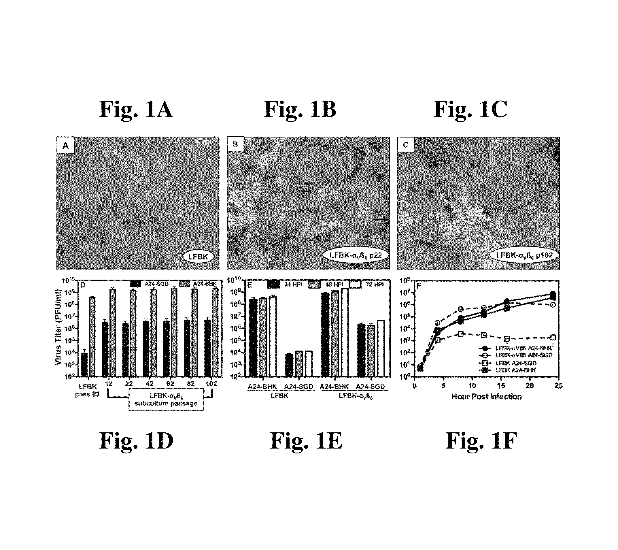
[0056]LFBK cells were propagated in DMEM supplemented with 10% fetal bovine serum and antibiotics as previously described (Piccone et al. 2009. J. Virol. 83:6681-6688) and used for these experiments between passages 64 and 71. LFBK-αVβ6 cells were propagated in the same manner as LFBK cells and used at the passage indicated in each experiment. BHK cells, used between passages 62 and 67, were propagated in MEM supplemented with 10% calf serum, 10% tryptose phosphate broth and antibiotics. Primary lamb kidney (LK) cells, generously supplied by the APHIS Diagnostic Service Section at PIADC, were propagated in DMEM supplemented with 10% fetal bovine serum, antibiotics and sodium pyruvate and used directly from cryovials or at passage 1 as indicated. IB-RS-2 cells were used between passage 129 and 137 and MVPK cells were used between passages 38 and 41; both of these cell lines were propagated in MEM supplemented with...

example 2

Susceptibility of LFBK-αVβ6 Cells to Animal-Derived FMDV of all Serotypes

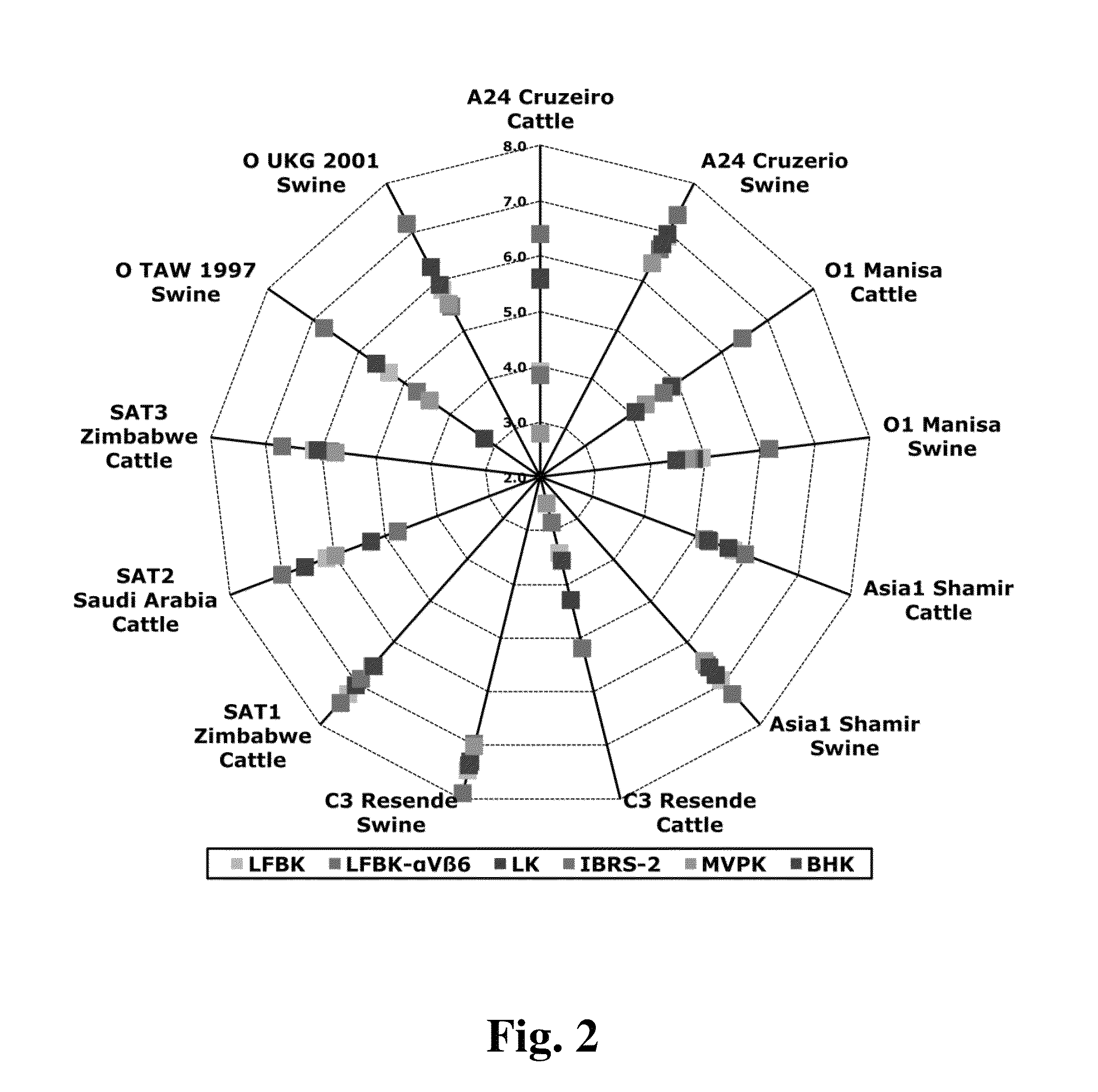
[0065]In order to determine the susceptibility of LFBK-αVβ6 cells to animal-derived FMDV strains and compare with that of LFBK, primary lamb kidney (LK), BHK, and two swine kidney cells lines (IB-RS-2 and MVPK), each cell type was infected with serial dilutions of infected tissue macerates or vesicular fluid obtained from animals experimentally infected with each of the FMDV serotypes. The viruses used in this experiment are listed in Table 1.

[0066]

TABLE 1Animal-derived viruses used in this study.SampleVirusSpeciesSample TypeA24 CruzeiroswineVesicular Fluid (VF)A24 CruzeirobovinePool of Tongue Tissue Homogenateand Vesicular FluidO1 ManisabovineVesicular FluidO1 ManisaswineVesicular FluidAsia1 ShamirbovineTongue Tissue HomogenateAsia1 ShamirswineVesicular FluidC3 ResendeswineVesicular FluidC3 ResendebovineTongue Tissue HomogenateO Taiwan 1997swineVesicular FluidO / TAW / 35 / 1997O UKGswineVesicular FluidUKG / 35 / 2001SA...

example 3

Detection of FMDV in Diagnostic Tissue Samples

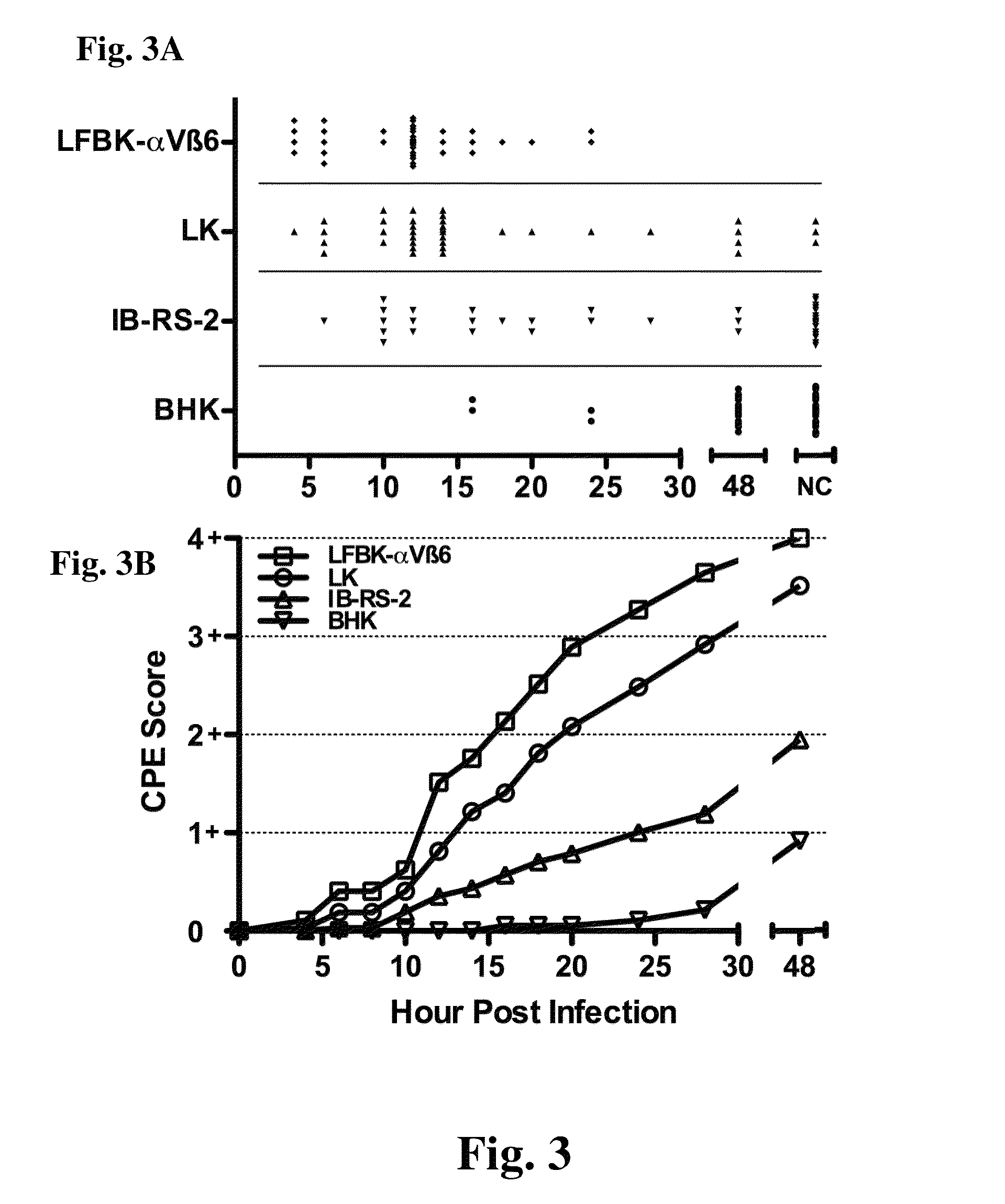
[0070]LK cells, BHK, IB-RS-2 and LFBK-αVβ6 were seeded onto 48-well cell culture plates 48 hours prior to infection. LK cells were seeded directly from storage cryovials. LFBK-αVβ6 cells were seeded at passage 32, IB-RS-2 at passage 136 and BHK-21 at passage 66. Diagnostic lesion tissues were disrupted and virus isolated after centrifugation through a Spin-X purification column (Costar) as described in (Pacheco et al. 2010, supra). 100 μl of a 1:10 dilution of these tissue macerates were used to infect each cell type for 1 hour at 37° C. After adsorption, 400 μl of growth media was added to each well and the plates were incubated at 37° C. Starting at 4 HPI, visual evidence of cytopathic effects was recorded every 2 hours until 20 HPI, then at 24, 28 and 48 HPI. At 48 HPI, all wells were fixed with 50% acetone:50% methanol and wells negative for cytopathic effects were immunohistochemically stained with a monoclonal antibody to FMDV 3D p...


PUM


Abstract

Foot and out disease virus (FMDV) is worldwide problem. Rapid isolation, serotyping and vaccine matching of FMDV from infected animals is critical to enable the implementation of effective vaccination programs and to stop the spread of infection during outbreaks. Current virus isolation protocols use primary cells, known to be susceptible to FMDV, or baby hamster kidney cells (BHK-21) and other cell lines that are not highly sensitive to some strains of FMDV. The αVβ6 integrin is a principal receptor for FMDV. We therefore transduced the porcine kidney cell line, LFBK, to stably express both the αV and β6 bovine integrin subunits. The LFBK-αVβ6 cell line showed both β6 expression and enhanced susceptibility to FMDV infection for at least 100 cell passages. LFBK-αVβ6 cells are highly sensitive for detecting all serotypes of FMDV from experimentally infected animals, including the porcinophillic strain O / TAW / 97 and are thus a sensitive tool for FMDV isolation.

Description

BACKGROUND OF THE INVENTION[0001]1. Field of the Invention[0002]The invention relates to a transgenic cell line which has been engineered to constitutively express the αVβ6 integrin receptor, a principal bovine receptor for Foot and Mouth Disease Virus (FMDV). In particular, the invention relates to the transgenic fetal porcine kidney cell line, LFBK αvβ6, which is useful for the rapid and sensitive detection and identification of FMDV in diagnostic settings and also to identify serotypes and subtypes thereby facilitating vaccine selection procedures. LFBK αvβ6 is highly sensitive and permissive to infection by animal-derived FMDV from all seven serotypes in cell culture. The invention further relates to the transgenic LFBK αvβ6 cells for detection of FMDV from field samples.[0003]2. Description of the Relevant Art[0004]Foot and mouth disease virus (FMDV) is a severe economic concern for meat producing nations since the trade of animal products is prohibited from countries where the...

Claims


Application Information

Patent Timeline
IPC IPC(8): C12N15/85G01N33/53C12N5/071
CPCC12N5/0684C12N2510/00C07K14/70546G01N33/56983C12N2760/20011C12N2760/20051C12N2770/16011C12N2770/16051C12N2770/32111C12N2770/32151C12N2770/32311C12N2770/32351C12N2710/24251C12N2710/24211C12N2770/32152C12Q1/04
Inventor LAROCCO, MICHAEL ABAXT, BARRYDUQUE, HERNANDO
Owner UNITED STATES OF AMERICA
Learn More