
Carbon nanotube anchor for mass spectrometer

a carbon nanotube and mass spectrometer technology, applied in the field of mass spectrometer sample anchors, can solve the problems of limiting the total achievable sensitivity, and achieve the effect of preventing supersaturation of the solution and reducing deposition on the area

Inactive Publication Date: 2013-12-03
UNIV OF SOUTH FLORIDA
View PDF16 Cites 5 Cited by
  • Summary
  • Abstract
  • Description
  • Claims
  • Application Information

AI Technical Summary

Benefits of technology

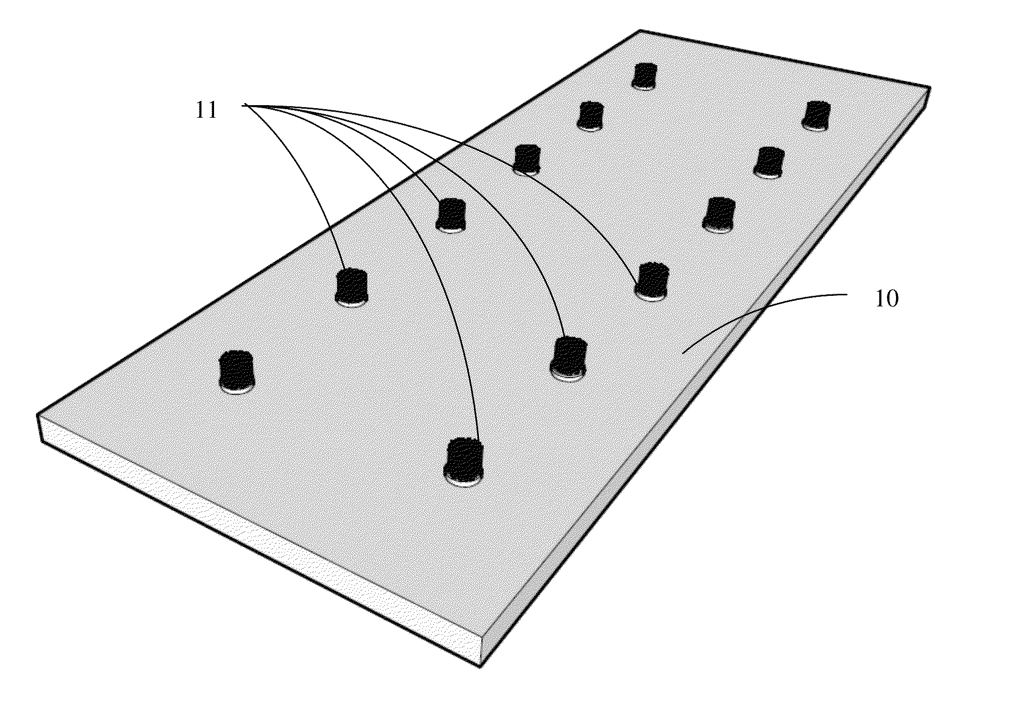
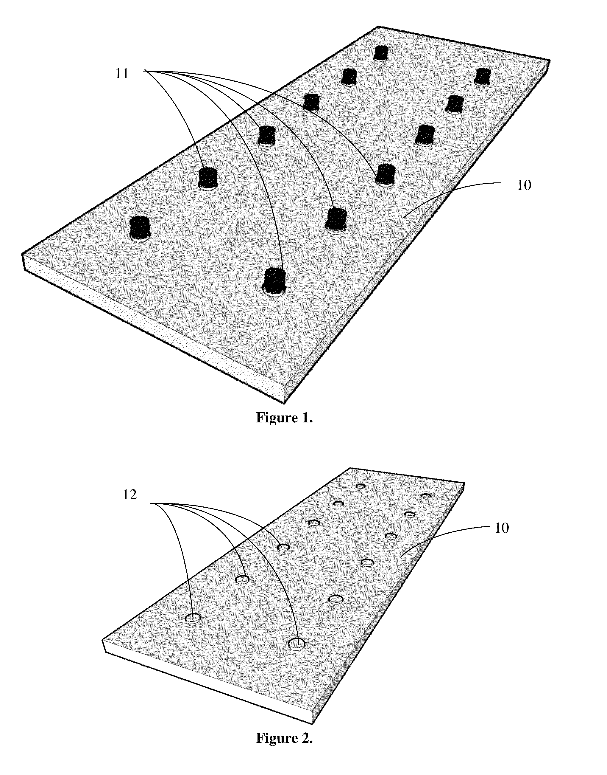
The carbon nanotube spots on the nanotube array act as nucleation centers, preventing solute molecules from clumping together and forming larger supersaturated areas. This reduces deposition on areas surrounding the nanotube spots and improves the accuracy and accuracy of the analysis. Other materials with a similar surface morphology can also be used for this purpose.

Problems solved by technology

This creates a supersaturation situation for the matrix molecules, causing them to precipitate on the area surrounding the anchor spot since the drop is still relatively large at that point, while in the same time collapsing due to the increased concentration.
Hence most of the analyte is never analyzed by the mass spectrometer, limiting the total achievable sensitivity.

Method used


Image

  • Carbon nanotube anchor for mass spectrometer
  • Carbon nanotube anchor for mass spectrometer
  • Carbon nanotube anchor for mass spectrometer

Examples


example 1

[0034]Prior to work with nonpolar matrices, such as HCCA, 3-HPA (water soluble) matrix was tested on a carbon nanotube enhanced substrate. A concentrating effect was witnessed with the carbon nanotube substrate, however it was similar to what can be observed using commercial anchor plates.

example 2

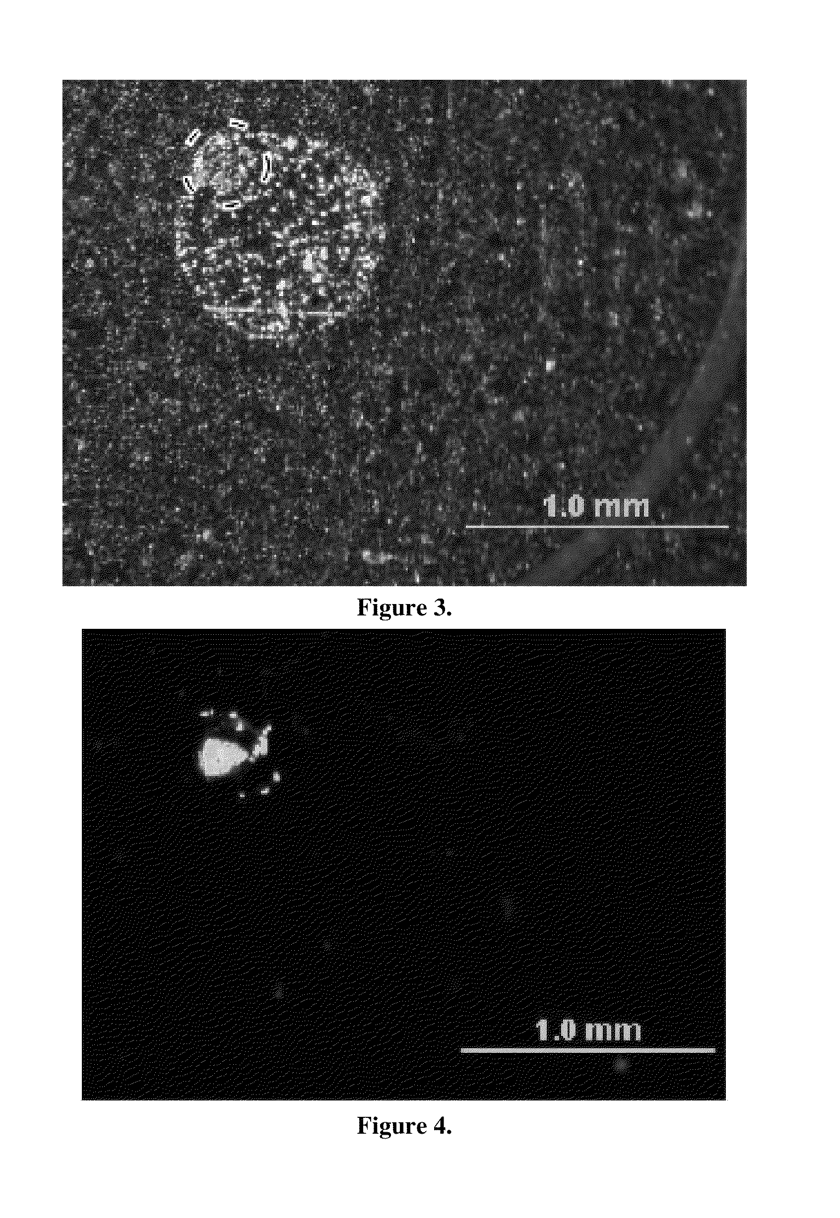
[0035]Analyte / matrix crystallization was compared on traditional target supports versus carbon nanotube-seeded target supports. 0.2 μL aliquots of 250 fmol / μL Mariner CALMIX 1 peptide standard solution (des-Arginine-Bradykinin, Angiotensin I, Glu-Fibrinopeptide, and Adrenocorticotropic Hormone (ACTH); Applied Biosystems (Foster City, Calif.)) mixed in a 3 mg / ml HCCA matrix compound was dropped onto a conventional Bruker “anchor plate” and allowed to evaporate. SEM images of the resulting solid matrix / analyte deposits display a diffuse 200 μm diameter anchor spot residue, seen in FIG. 3. Matrix concentrations were varied proportionally with the analyte, from 0.3 mg / mL to 0.006 mg / mL HCCA, to keep the matrix to analyte ratio constant. The matrix / analyte deposit is considerably larger (˜0.8 mm diameter) than the anchor spot (circled) on the Bruker target support. It was also noted that this large matrix / analyte deposit was similar in magnitude to previous studies on MALDI matrix / analyt...

example 3

[0038]Carbon nanotube anchor spots were found to vastly improve MALDI-TOF-MS signal-to-noise ratios. An Applied Biosystems Voyager DE STR MALDI-TOF and nitrogen laser at 20 Hz firing frequency with 400 micron fiber coupling were used for these experiments. Glu-Fibrinopeptide samples were then digested and mixed in a 2.5 mg / ml HCCA matrix. 0.2 μL aliquots of 250 fmol / μL were applied to either the traditional Bruker support or the carbon nanotube target support and ionized by a laser and run through a mass spectrometer with the operating laser intensity set slightly above threshold levels, and an acquisition of 250 shots per spectrum. The samples were analyzed on single positions on the target support using a delayed-extraction mode (extraction delay 200 nanoseconds). Signal-to-noise ratios for 1570 m / z peak in a variety of analyte concentrations were recorded, as seen in FIG. 6. The nanotube-based anchor spot samples display at least a 3-5× better signal than the traditional Bruker s...


PUM

PropertyMeasurementUnit
Lengthaaaaaaaaaa
Lengthaaaaaaaaaa
Areaaaaaaaaaaa

Abstract

This invention enables a sensitivity enhancement in the detection of molecular compounds. A mass spectrometry analyte support with nanotube anchors are used to concentrate MALDI samples, specifically samples prepared with water-insoluble matrix compounds, on the anchor spot. The surface structure is established through patterned carbon nanotube anchor growth, providing a nucleation center for analyte and reducing sample precipitation on the surrounding MALDI wafer. Also disclosed is a method of creating a mass spectrometry support using patterned metal catalyst to grow carbon nanotubes. The carbon nanotubes enhance nucleation on specific areas of a sample plate to concentrate analyte / matrix deposit during droplet evaporation.

Description

CROSS REFERENCE TO RELATED APPLICATION[0001]This application claims priority to currently pending U.S. Provisional Patent Application No. 61 / 033,909, entitled “Carbon Nanotube Anchor for Mass Spectrometer”, filed on Mar. 5, 2008, the contents of which are herein incorporated by reference.FIELD OF INVENTION[0002]This invention relates to mass spectrometer sample anchors. Specifically, the invention is a carbon nanotube anchor plate for water insoluble samples in matrix assisted laser desorption ionization mass spectrometers.BACKGROUND OF THE INVENTION[0003]Matrix assisted laser desorption ionization mass spectrometry (MALDI-MS) is a technique used for the quantification and detection of bio-molecules and other macro-molecular substances for applications ranging from proteomics and cancer early detection to forensic investigations. Especially for proteomics and cancer research, sensitivity and analyte concentration are essential for successful measurements, since the analyte is often ...

Claims


Application Information

Patent Timeline
IPC IPC(8): G21K5/04
CPCH01J49/0418G01N2560/00C23C16/455H01J49/164
Inventor SCHLAF, RUDIGERSCHUMACHER, JOSHUA
Owner UNIV OF SOUTH FLORIDA
Learn More