
Alginate polyelectrolyte encapsulation of embryonic stem cells

a technology of embryonic stem cells and alginate polyelectrolyte, which is applied in the field of alginate polyelectrolyte microencapsulation of cells, can solve the problems of limited development of effective tissue-engineered artificial liver devices, generated from autogenic or allogeneic cells, and prior techniques that have been limited in a number of ways

Active Publication Date: 2013-05-07
RUTGERS THE STATE UNIV
View PDF6 Cites 2 Cited by
  • Summary
  • Abstract
  • Description
  • Claims
  • Application Information

AI Technical Summary

Benefits of technology

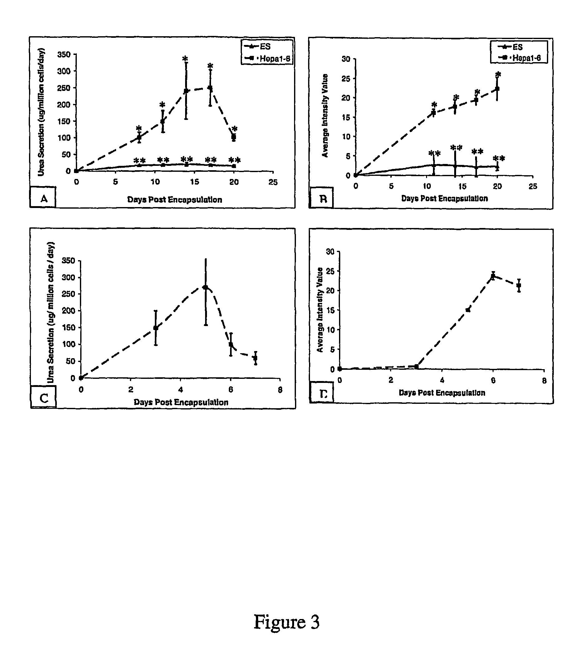
This approach enables scalable, controlled differentiation of embryonic stem cells into functional hepatocytes with high viability and functional expression, maintaining over 90% cell viability and promoting hepatocyte-specific functions like urea secretion and albumin production, without the need for cumbersome embryoid body formation or growth factors.

Problems solved by technology

The development of effective tissue-engineered artificial liver devices, generated from autogenic or allogeneic cells, has been limited by the absence of large numbers of mature, functional hepatocytes.
However, the development of these devices has similarly been limited by the absence of large numbers of mature functional hepatocytes.
Although several publications have described techniques to effectively differentiate stem cells into hepatocytes (6), these prior techniques have been limited in a number of ways.
Such limitations include the absence of large scale processing considerations, incomplete downstream enrichment techniques, and ineffective long term maintenance of functional hepatocytes.
A disadvantage of this approach was that it was necessary to release the compact colonies of ES cells from the microbeads before differentiation in adhesion cultures could be induced.
Before the present invention, approaches to differentiate ES cells into specific cell types within alginate beads had limited success and had practical limitations.
For example, previously, differentiation of ES colonies within 1.1% alginate microbeads, which had weak mechanical resistance, could only be promoted by supplementation of the cell media with retinoic acid, and required the cumbersome formation of embryoid bodies.

Method used


Image

  • Alginate polyelectrolyte encapsulation of embryonic stem cells
  • Alginate polyelectrolyte encapsulation of embryonic stem cells
  • Alginate polyelectrolyte encapsulation of embryonic stem cells

Examples


example 1

Materials and Methods

Cell Culture

[0085]All cell cultures were incubated in a humidified 37° C., 5% CO2 environment. The ES cell line D3 (ATCC, Manassas, Va.) was maintained in an undifferentiated state in T-75 gelatin-coated flasks (Biocoat, BD-Biosciences, Bedford, Mass.) in Knockout Dulbecco's modified Eagles medium (Gibco, Grand Island, N.Y.) containing 15% knockout serum (Gibco), 4 mM L-glutamine (Gibco), 100 U / ml penicillin (Gibco), 100 U / ml streptomycin (Gibco), 10 μg / ml gentamicin (Gibco), 1000 U / ml ESGRO (Chemicon, Temecula, Calif.), 0.1 mM 2-mercaptoethanol (Sigma-Aldrich, St. Louis, Mo.). ESGRO contains leukemia inhibitory factor (LIF) which prevents embryonic stem cell differentiation. Every 2 days, media was aspirated and replaced with fresh media. Cultures were split and passaged every 6 days, following media aspiration, washing with 6 ml of phosphate buffered solution (PBS) (Gibco). Cells were detached following incubation with 3 ml of trypsin (Gibco) for three minutes...

example 2

Encapsulation System

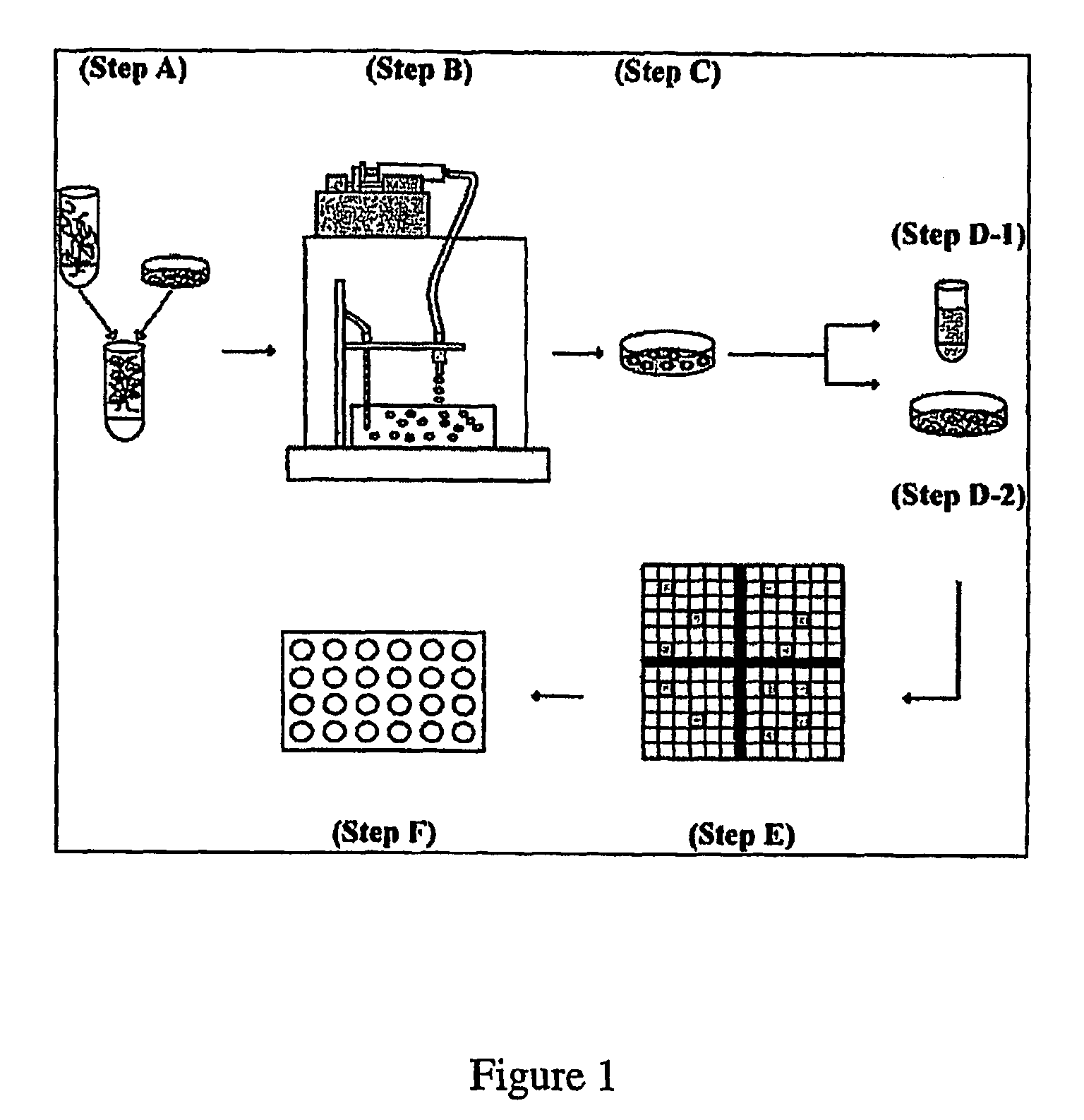
[0096]An alginate-PLL murine embryonic stem cell encapsulation system was developed in order to investigate the feasibility of regulating hepatocyte differentiation within a controlled microenvironment and recovering differentiated cells from intracapsular culture. One embodiment of the encapsulation system is shown in FIG. 1. Briefly, embryonic stem cells were detached from culture dishes and mixed with various concentrations of dissolved alginate (Step A). The alginate cell suspension mixture was transferred to a 10 ml syringe within an electrostatic bead generator (Step B), which was used to control bead size through an applied voltage (6.5 kV). The alginate cell suspension was extruded into a bath of 100 mM calcium chloride and the beads were subsequently coated in a poly-L-lysine solution to increase bead durability. The encapsulated cell population was then cultured for 20 days, either in the presence or absence of LIF (Step C). On days 8, 11, 14, 17 and 20...

example 3

Assessment of Biocompatibility

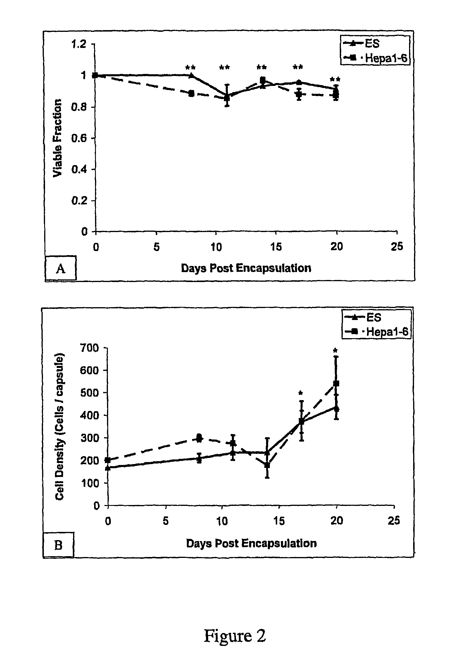
[0097]Initial experiments were designed to determine the biocompatibility of alginate-PLL microencapsulation using both Hepa1-6 murine hepatoma cells and undifferentiated murine embryonic D3 cells. Immediately following encapsulation, beads were stained with calcein / ethidium homodimer, imaged, and the number of viable cells within the beads determined. On average, the percent viability was reduced minimally from 98% to 95%, indicating that the encapsulation process caused little cell damage. The effect of depolymerization and recovery on cell viability was also assessed.

[0098]The results are shown in FIG. 2 (A and B). In these experiments, ES cells were encapsulated in 2.0% w / v alginate, at a cell seeding density of 5×10⁶ cells / ml, and cultured in Knockout DMEM containing ESGRO to prevent differentiation; Hepa 1-6 cells were encapsulated in 2.0 w / v alginate, at a cell seeding density of 5×10⁶ cells / ml, and cultured in DMEM. In FIG. 2, each data point re...


PUM

PropertyMeasurementUnit
bead sizeaaaaaaaaaa
voltageaaaaaaaaaa
sizeaaaaaaaaaa

Abstract

Alginate polyelectrolyte encapsulation is used for the controlled differentiation of embryonic stem cells. An isolated cell population is provided. The cell population includes a single cell suspension of ES cells encapsulated within an alginate polyelectrolyte microenvironment. The encapsulated ES cells are capable of differentiating within said microenvironment into hepatocyte lineage cells in the absence of embryoid body intermediates or growth factor supplementation.

Description

FIELD OF THE INVENTION[0001]The present invention relates to alginate polyelectrolyte microencapsulation of cells. In particular, the invention is directed to the use of alginate poly-L-lysine microencapsulation in the controlled differentiation of embryonic stem cells.BACKGROUND[0002]A stem cell is a cell that has the ability to replicate itself for indefinite periods and to give rise (differentiate), under suitable conditions, to the many different cell types that make up an organism. That is, stem cells have the ability to develop into mature cells that have characteristic morphology and specialized functions, such as liver cells, heart cells, skin cells, nerve cells, etc.[0003]Embryonic stem (ES) cells are able to differentiate into virtually all cell and tissue types of an organismin vivo as well as in vitro. Unlike other stem cells, they can differentiate into cells that are derived from all three primary germ layers: the ectoderm, mesoderm or endoderm. Each cell type and tis...

Claims


Application Information

Patent Timeline
Patent Type & Authority Patents(United States)
IPC IPC(8): C12N15/00A61K35/545C12N5/071
CPCA61K35/545C12N5/067C12N2506/02C12N2533/32C12N2533/74
Inventor MAGUIRE, TIMSCHLOSS, RENE S.YARMUSH, MARTIN L.
Owner RUTGERS THE STATE UNIV
Learn More