Eureka AIR delivers breakthrough ideas for toughest innovation challenges, trusted by R&D personnel around the world.

Systems, processes and software arrangements for evaluating information associated with an anatomical structure by an optical coherence ranging technique

an anatomical structure and optical coherence ranging technology, applied in the field of systems, processes and software arrangements for evaluating information associated with anatomical structures by optical coherence ranging technique, can solve the problems of high scattering, increased costs to the health care system and to the individual patient, and differences between the data obtained by these prior art methods and the medical standards of care for diagnosis

Inactive Publication Date: 2013-01-08
THE GENERAL HOSPITAL CORP
View PDF590 Cites 56 Cited by
  • Summary
  • Abstract
  • Description
  • Claims
  • Application Information

AI Technical Summary

Benefits of technology

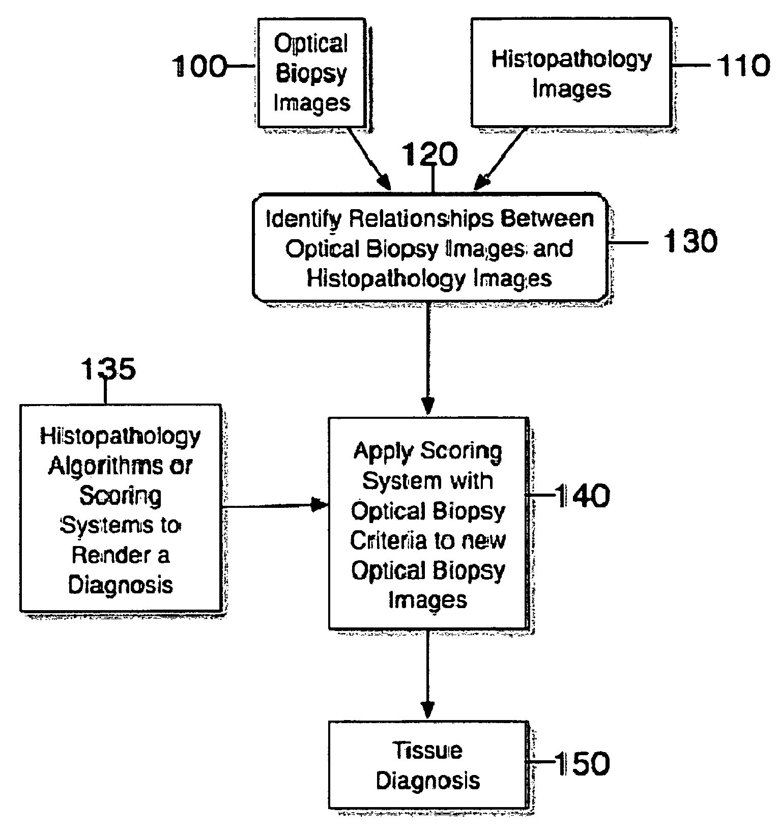
[0026]These and other objects can be achieved by providing an exemplary embodiment of the system, process and software arrangement according to the present invention for obtaining histopathologic diagnoses from optical biopsy images. For example, it is possible to determine a relationship between the properties of the optical biopsy images and images / data utilized in the practice of medicine and pathology to provide a diagnosis. These properties may include, but not limited to a resolution, modes of contrast, spatial and structural features, etc. For example, the resolution of OCT images may be similar to the resolution obtained by a microscope view at low power. Therefore, exemplary architectural features visualized by the OCT systems and methods may be compared to architectural features seen by the conventional histopathology. The contrast in the OCT images may be analogous or similar to conventional histopathologic H&E stains in that high scattering, which may occur at regions of high nuclear content that may be similar or analogous to the basophilic stain of hematoxylin. Furthermore, collagen may have a high scattering observed by the OCT systems and methods, which can be related to linear eosinophilic staining of the lamina propria and submucosa seen in histopathology. These exemplary features of the OCT techniques, systems and method assist with a determination of an analogy between architectural structures seen by this optical biopsy technique and by a low power view of microscopic tissue typically viewed by pathologists.
[0027]According to one exemplary embodiment of the present invention, when a relationship is established between the optical biopsy images and histopathology images, criteria and methods used by pathologists to interpret H&E stained slides can be modified on the basis of the comparison between image features and characteristics. Scoring systems and techniques based on these criteria and methods can then be modified and applied to the optical biopsy images themselves. In this manner, a diagnosis can be obtained from the optical biopsy images that are related to conventional histopathologic diagnoses. Advantages of this exemplary embodiment of the system, process, software arrangement and technique can include an ability to utilize prior information obtained from histopathologic correlations between morphology and outcome. Furthermore, since the histopathologic correlations have been formed over decades, the optical biopsy criteria can be similarly reliable for predicting the patient outcome. This can result in a tissue diagnosis from these non- or minimally-invasive methods.

Problems solved by technology

While these conventional devices can provide information that is related to disease, there are differences between the data obtained by these prior art methods and the medical standards of care for diagnosis.
Such increases may incur significant costs to the health care system and to the individual patient.
Furthermore, certain methods which do not utilize endoscopy can potentially be conducted at a lower cost, partially alleviating the financial burden of comprehensive screening on the health care system.
The accuracy of surveillance endoscopy, however, may be limited by a sampling error, as discussed in G. S. Dulai, “Surveying the case for surveillance,” Gastroenterology, 2002, Vol. 122(3), pp.
Such increases can incur significant costs to the health care system and to the individual patient.

Method used


Image

  • Systems, processes and software arrangements for evaluating information associated with an anatomical structure by an optical coherence ranging technique
  • Systems, processes and software arrangements for evaluating information associated with an anatomical structure by an optical coherence ranging technique
  • Systems, processes and software arrangements for evaluating information associated with an anatomical structure by an optical coherence ranging technique

Examples


example 1

Determination of SIM at SCJ from OCT Images

[0058]i. Exemplary Design

[0059]An exemplary study for the exemplary embodiments of the present invention was a blinded, prospective trial. Its primary objective was to identify OCT image features for differentiating intestinal metaplasia at the SCJ. Patients undergoing a routine outpatient upper endoscopy were requested to participate in the study. OCT images of the SCJ were obtained during the endoscopy procedure. Two pathologists reviewed each biopsy specimen, and noted the presence of the following tissue types: gastric or oxyntic cardia, squamous mucosa, pancreatic metaplasia. The existence of intestinal metaplasia was noted by the presence of goblet cells. Image features of intestinal metaplasia were determined by creating and reviewing an OCT atlas “training set”, containing biopsy-correlated images of known tissue types. These features were then prospectively applied to a “validation set” of unknown tissue types. The sensitivity, spe...

example 2

Identifying High Grade Dysplasia and Intramucosal Carcinoma in OCT Images of SIM

[0082]i Exemplary Study Design

[0083]The exemplary study performed was a blinded, prospective trial. Recruited subjects were patients with BE undergoing routine endoscopic surveillance or confirmatory biopsies for IMC or HGD. OCT images of the Barrett's epithelium were obtained during endoscopy. Biopsy correlated OCT images of the esophagus were viewed and scored by a reader blinded to the tissue diagnosis. For each image the score for surface maturation and gland architecture were summed to establish a “dysplasia index”. Each biopsy specimen was independently reviewed, and a consensus diagnosis was rendered.

[0084]ii Exemplary OCT System

[0085]The exemplary OCT device which can be utilized for the exemplary embodiment of the present invention and used in the study is described in J. M. Poneros et al., “Diagnosis of specialized intestinal metaplasia by optical coherence tomography,” Gastroenterology, 2001, ...


PUM


Abstract

Software systems, arrangements and processes for evaluating an image associated with at least one portion of an anatomical structure are provided. For example, first information associated with the at least one portion of the anatomical structure second information associated with the at least one portion of the anatomical structure can be received. Third information can be generated by determining a relationship between the first information and the second information. Further, the image can be evaluated using a predetermined pathological scoring criteria and the third information.

Description

CROSS-REFERENCE TO RELATED APPLICATION(S)[0001]This application is based upon and claims the benefit of priority from U.S. patent application Ser. No. 60 / 676,362, filed Apr. 28, 2005, the entire disclosure of which is incorporated herein by reference.STATEMENT REGARDING FEDERALLY SPONSORED RESEARCH[0002]The invention was made with the U.S. Government support under Contract No. RO1 CA 103767 awarded by the National Institute of Health. Thus, the U.S. Government has certain rights in the invention.FIELD OF THE INVENTION[0003]The present invention relates to systems, processes and software arrangements for evaluating information associated with an anatomical structure by optical coherence ranging evaluating information by an optical coherence ranging technique, and for example for interpreting microscopic images obtained from living subjects.BACKGROUND INFORMATION[0004]A variety of different optical biopsy techniques have been described and developed for a non-invasive diagnosis of dis...

Claims


Application Information

Patent Timeline
Patent Type & Authority Patents(United States)
IPC IPC(8): G06K9/00G16H30/20G16H70/60
CPCA61B10/02G06T11/003A61B5/0033G06T7/0014A61B2010/0003A61B2017/00827G06F19/321G06T2207/20081G06T2207/30004G16H70/60G16H30/20H04N17/00G06T7/00A61B5/0066G06T2207/10101G06T2207/30096
Inventor TEARNEY, GUILLERMO J.BOUMA, BRETT EUGENEEVANS, JOHN A.
Owner THE GENERAL HOSPITAL CORP
Why Eureka
Eureka Blog
Learn More