Anti-5T4 antibodies and uses thereof
a technology of anti-5t4 antibodies and conjugates, which is applied in the direction of antibody medical ingredients, drug compositions, peptide sources, etc., can solve the problems of limited human immunotherapy and rapid antibody clearance, and achieve the effect of enhancing drug incorporation
- Summary
- Abstract
- Description
- Claims
- Application Information
AI Technical Summary
Benefits of technology
Problems solved by technology
Method used
Image
Examples
example 1
Murine Anti-5T4 Antibodies
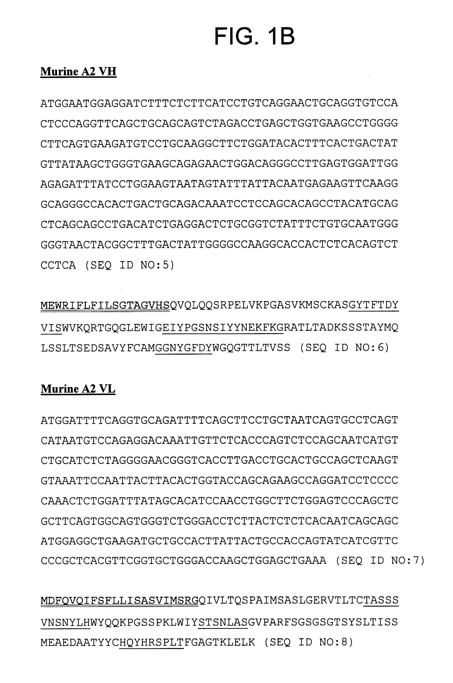
[0222]Anti-5T4 antibodies were prepared in mice using human 5T4 antigen and standard methods for immunization. Hybridoma cell lines producing the A1, A2, and A3 antibodies were produced by fusion of individual B cells with myeloma cells.
[0223]The A1, A2, and A3 anti-5T4 antibody heavy chain and light chain variable regions were cloned using the SMART® cDNA synthesis system (Clontech Laboratories Inc. of Mountain View, Calif.) followed by PCR amplification. The cDNA was synthesized from 1 μg total RNA isolated from A1, A2, or A3 hybridoma cells, using oligo(dT) and the SMART® IIA oligo (Clontech Laboratories Inc.) with POWERSCRIPT™ reverse transcriptase (Clontech Laboratories Inc.). The cDNA was then amplified by PCR using a primer which anneals to the SMART® IIA oligo sequence and mouse constant region specific primer (mouse Kappa for the light chain, mouse IgG2a for the A1 heavy chain, mouse IgG2b for the A2 heavy chain, and mouse IgG1 for the A3 heavy cha...
example 2
Binding Specificity and Affinity of Murine Anti-5T4 Antibodies
[0228]To assess the binding specificity and affinity of the A1, A2, and A3 antibodies, BIACORE® analysis was performed using human 5T4 antigen immobilized on a CM5 chip. BIACORE® technology utilizes changes in the refractive index at the surface layer upon binding of the antibody to the 5T4 antigen immobilized on the layer. Binding is detected by surface plasmon resonance (SPR) of laser light refracting from the surface. Analysis of the signal kinetics on rate and off rate allows discrimination between non-specific and specific interactions. The H8 anti-5T4 antibody was used as a control. H8 is a hybridoma-generated monoclonal mouse IgG1 antibody described in PCT International Publication No. WO 98 / 55607 and in Forsberg et al. (1997) J. Biol. Chem. 272(19):124430-12436.
[0229]
TABLE 3Results of BIACORE ® AssayAntibodyKD (M)KA (1 / M)kd (1 / s)ka (1 / Ms)H84.1 × 10−102.5 × 1095.1 × 10−51.3 × 10⁵A16.4 × 10−101.6 × 1091.3 × 10−42.0 ...
example 3
Internalization of Murine Anti-5T4 Antibodies by 5T4-Expressing Cells
[0235]To assess internalization of antibodies upon binding to 5T4 antigen, the amount of H8 and A1 antibodies detected at the cell surface versus in the supernatant was determined as a function of time. Non-enzymatically dissociated MDAMB435 / 5T4 cells (human breast cancer cells) were exposed to anti-5T4 antibodies for 1 hour at 4° C. Cells were washed and incubated in media at 37° C. for 4 hours or 24 hours. The amount of antibody bound to cellular membranes versus unbound antibody (i.e., presence in the supernatant) was determined using FACS. The disappearance of 5T4 antibodies from the surface of MDAMB435 / 5T4 cells demonstrates modulation of the 5T4 antigen / antibody complex at the cell surface, which may indicate internalization and / or dissociation. See FIGS. 4A-4C.
PUM
 Abstract
Description
Claims
Application Information
 - R&D
- Intellectual Property
- Life Sciences
- Materials
- Tech Scout
- Unparalleled Data Quality
- Higher Quality Content
- 60% Fewer Hallucinations
Browse by: Latest US Patents, China's latest patents, Technical Efficacy Thesaurus, Application Domain, Technology Topic, Popular Technical Reports.
© 2025 PatSnap. All rights reserved.Legal|Privacy policy|Modern Slavery Act Transparency Statement|Sitemap|About US| Contact US: help@patsnap.com


