Time-dependent three-dimensional musculo-skeletal modeling based on dynamic surface measurements of bodies

a dynamic surface measurement and musculoskeletal technology, applied in the field of time-dependent three-dimensional musculoskeletal modeling based on dynamic surface measurements of bodies, can solve the problems of large number of people suffering from different musculoskeletal complaints, such as back pain or knee problems, and provide a non-contact technique for obtaining, so as to achieve short recording and analysis time, no preparation time, and the effect of efficient, stable and reliable devices

Active Publication Date: 2011-03-01
DIERS ENG
View PDF4 Cites 6 Cited by
  • Summary
  • Abstract
  • Description
  • Claims
  • Application Information

AI Technical Summary

Benefits of technology

[0024]It is an advantage of the invention that with the equipment it is possible to reconstruct kinematics, kinetics and dynamics of the musculo-skeletal system of the body, as well as to indicate and quantify pathological changes or abnormalities.
[0025]It is furthermore an advantage of the invention that no preparation time and only a short recording and analysing time are needed.
[0026]It is an advantage of the invention that it enables the full time-dependent three-dimensional measurement and functional analysis of the human body in a contact-free, non-invasive way, without the use of potentially harmful radiation. It is furthermore an advantage of the present invention that it allows the use of time-dependent three-dimensional images of the surface of part of a body for construction of a musculo-skeletal model.
[0027]It is an advantage of the present invention that, for the detection of time-dependent anatomical surface information on a series of time-dependent three-dimensional images of the surface, both detection points and characteristics of parts of the surface are used.
[0028]It is furthermore an advantage of the present invention that the time evolution of anatomical surface information and thus the characteristics of parts of the surface are used during construction of the musculo-skeletal model.
[0029]Although there has been constant improvement, change and evolution of devices in this field, the present concepts are believed to represent substantial new and novel improvements, including departures from prior practices, resulting in the provision of more efficient, stable and reliable devices of this nature.

Problems solved by technology

The amount of people suffering from different musculo-skeletal complaints, such as back pain or knee problems, is large.
Up to now, time-dependent three-dimensional recordings of movement under functional conditions are not possible with conventional techniques.
US 2002 / 0009222 does not provide a non-contact technique for obtaining information and furthermore does not use muscular modelling.
The application of landmark markers to the body is time consuming and must be tolerated by patients.
The document describes the construction of a model based on expensive techniques and involving potentially harmful techniques which require careful management.
Furthermore, the methods described in the above mentioned documents have the disadvantage that the computing power needed for time-dependent detection of landmarks on time-dependent images of body parts can be high.
The above mentioned documents furthermore have the disadvantage that a musculo-skeletal model only can be obtained based on images of the interior of the body parts.
Furthermore, the above mentioned documents have the disadvantage that the musculo-skeletal model obtained can lead to bio-mechanical inconsistent features and that the computing power for obtaining the musculo-skeletal model is large.
Due to the large computing power needed, the systems and methods for obtaining a musculo-skeletal model are tedious and labour-intensive.

Method used


Image

  • Time-dependent three-dimensional musculo-skeletal modeling based on dynamic surface measurements of bodies
  • Time-dependent three-dimensional musculo-skeletal modeling based on dynamic surface measurements of bodies
  • Time-dependent three-dimensional musculo-skeletal modeling based on dynamic surface measurements of bodies

Examples


example 1

Spine

[0101]The internal spine is reconstructed from the external spine using an anatomical formula, estimating the distance of the skin to the centre of a vertebral body, as illustrated on FIGS. 15 and 16. Spinal parameters (e.g. lumbar lordisis angle) can be monitored as a function of time in three dimensions. A detailed deduction of the anatomical formula is described by Drerup et al. in Clinical Biomechanics 9 p 28-36.

example 2

Shoulder

[0102]The shoulder is a complex joints and it is not possible to model it as a 3 degrees-of-freedom spherical joint. The skeletal model 240, as shown in FIG. 20, contains the following bones: the sternum, clavicula, scapula and humerus. Between the bones, three joints have been defined: the sternoclavicular joint, the acromioclavicular joint and the glenohumeral joint. These joints are modelled as three-degrees-of-freedom (DOFs) spherical joints. The scapulothoracic joint is modelled in such a way that the scapula is able to move freely with respect to the thoracic wall, in order to enable winging. The set of measured anatomical features is necessary and sufficient to define all DOF's of the system, and to build a personalised time-dependent three-dimensional model of the shoulder complex.

example 3

Pelvis

[0103]A personalised time-dependent three-dimensional model of the pelvis is reconstructed from the lower back surface and the position of the dimple points.

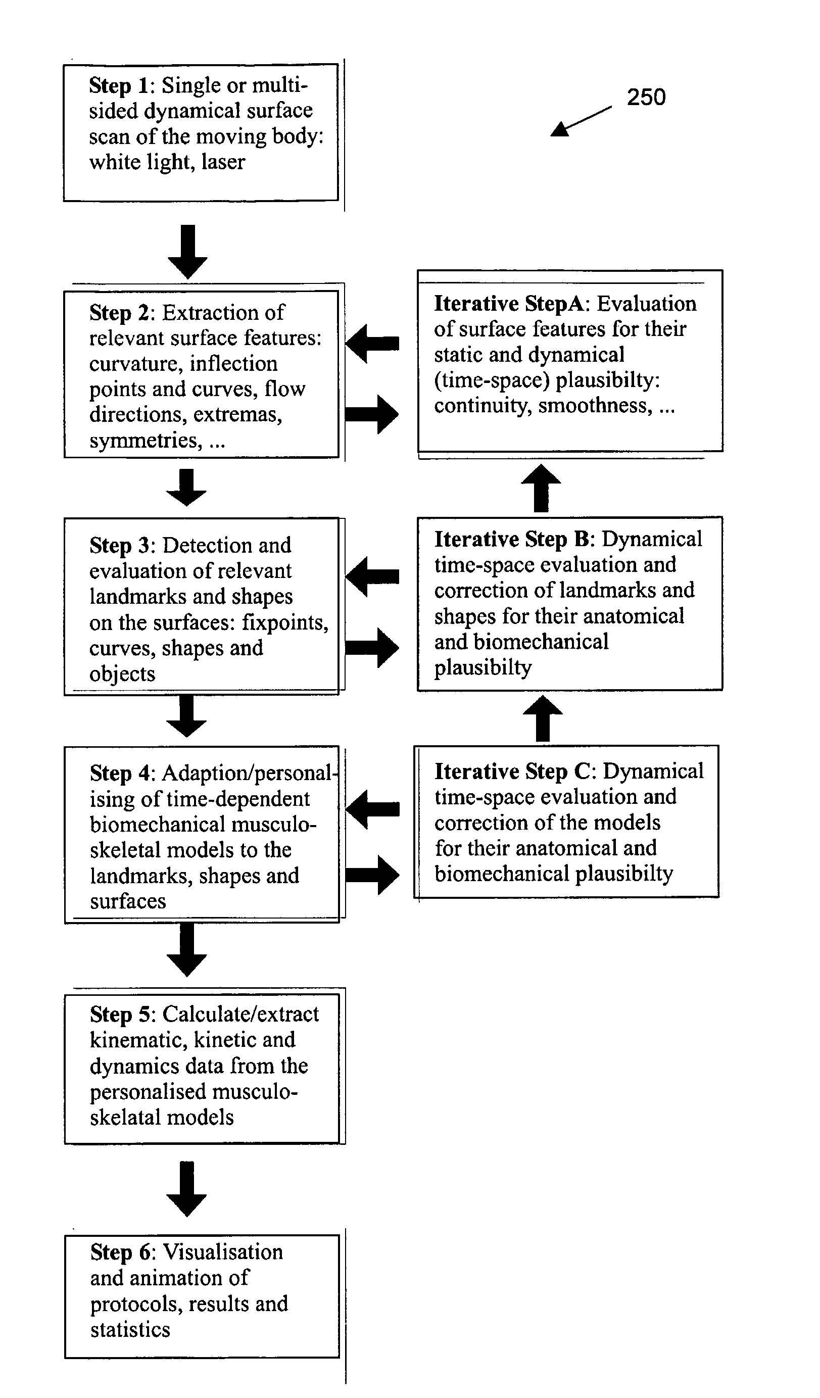
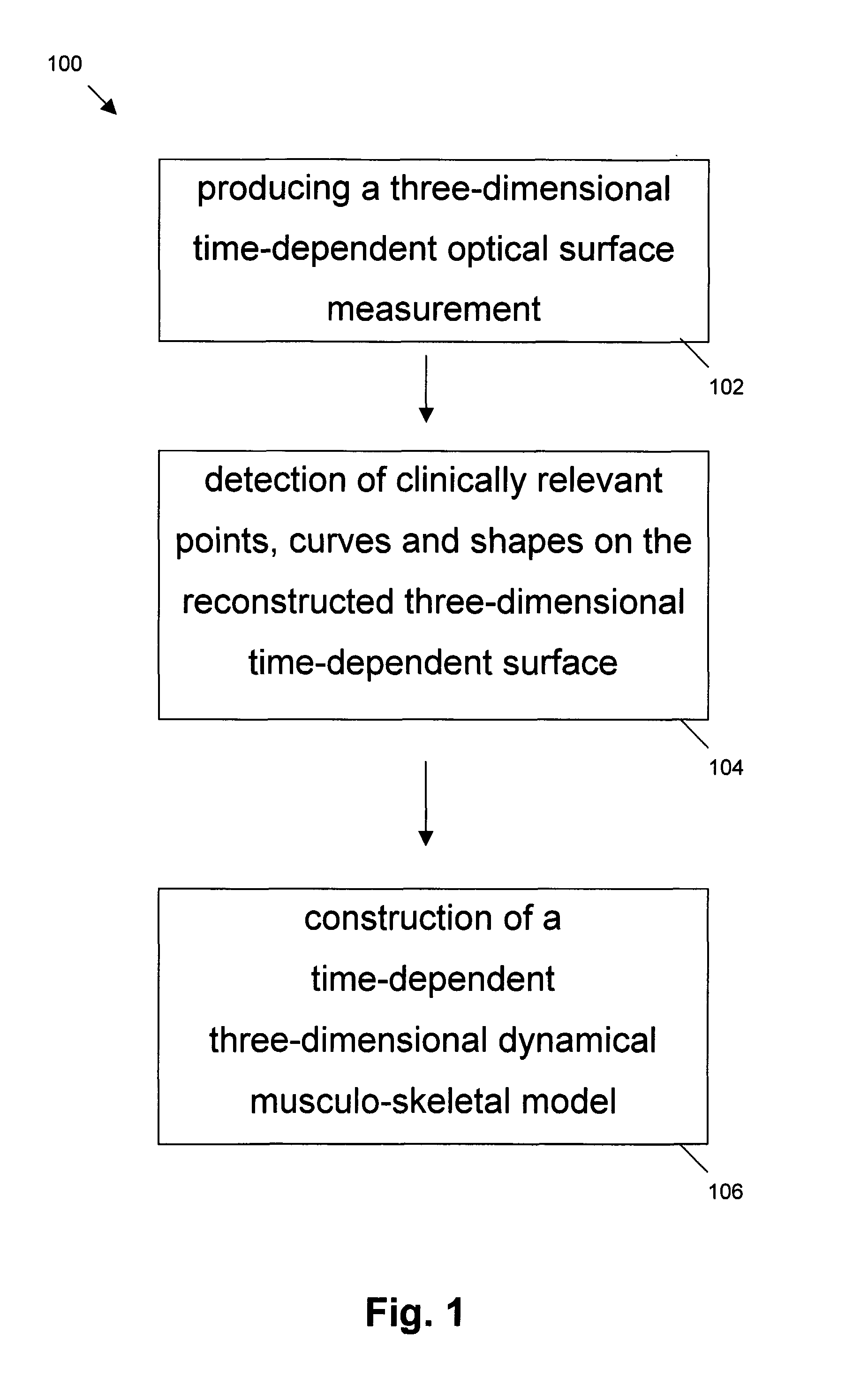
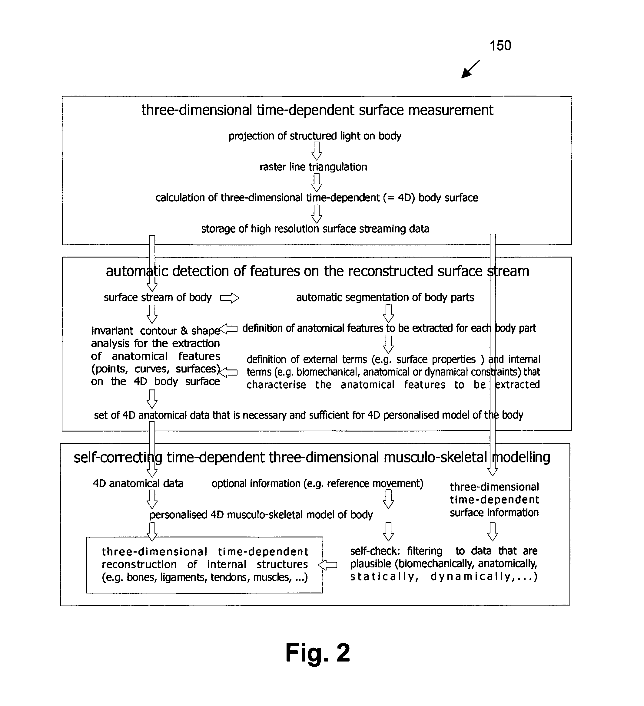
In conclusion, the method of the above described embodiment comprises the provision, e.g. grabbing, of surface images of a moving body by suitable scanning or projection equipment, e.g. optical equipment. Thereby either a one-sided surface (like human back, face, . . . ) or a multi-sided surface (legs, torso, . . . ) is recorded. The method furthermore comprises the reconstruction and mapping on a regular grid of the surface, thus giving a sequence of single static surfaces. On the regular surfaces, invariant features are calculated, like curvature and symmetry and invariant features are used for detecting anatomical landmarks and shapes on each of the static surface, like vertebra prominence, sacrum point (rima ani), left and right dimples, acromium, scapula, spinal symmetry line (processi spinosi). In the next step, the ...


PUM


Abstract

Active contour models and active shape models were developed for the detection of the kinematics landmarks on sequential back surface measurements. The anatomical landmarks correspond with the spinous process, the dimples of the posterior superior iliac spines (PSIS), the margo mediales and the elbow. Back surface curvatures are used as a basis to guide the ACM and ASM's towards interesting landmark features on the back surface. Geometrical bending and torsion costs, and the main modes of variation of the landmark points are added to the models in order to avoid unrealistic curve shapes from a biomechanical point of view. Reconstruction of the underlying skeletal structures is performed using the surface normals as approximations for skeletal rotations (e.g. axial vertebrae rotations, pelvic torsion, etc.) and anatomical formulas to estimate skeletal dimensions.

Description

CROSS-REFERENCE TO RELATED APPLICATIONS[0001]This application is the U.S. National Stage of International Application No. PCT / BE2005 / 000031, filed Feb. 25, 2005, which, in turn, claims the benefit of British Patent Application Serial No. 0404269.3, filed Feb. 26, 2004.TECHNICAL FIELD OF THE INVENTION[0002]The invention relates to a system and method for time-dependent three-dimensional measurement and functional analysis of bodies. In particular the invention relates to a system and method for obtaining a time-dependent and three-dimensional musculo-skeletal system for bodies or body parts. The invention furthermore relates to a system and method for quickly and accurately detecting landmarks on three-dimensional and time-dependent images of body parts and to a system and method for constructing a feasible or approximate musculo-skeletal model based on three-dimensional and time-dependent images of body parts.BACKGROUND OF THE INVENTION[0003]The amount of people suffering from diffe...

Claims


Application Information

Patent Timeline
Patent Type & Authority Patents(United States)
IPC IPC(8): G06K9/00A61B5/103G06T15/70G06T17/20
CPCG06T7/0012G06T7/2033G06T17/20A61B5/0064G06T2207/10016G06T2207/20101G06T2207/20116G06T2207/20124G06T2207/30012G06T2207/30196G06T2207/30204G06T7/246
Inventor HAEX, BART MARIA JOZEFVANDER SLOTEN, JOZEFDIERS, HELMUTHEITMANN, KJELL ROGER
Owner DIERS ENG
Why Eureka
Try Eureka