
Soft tissue processing

a soft tissue and processing technology, applied in the field of soft tissue processing, can solve the problems of narrow therapeutic window between adequate immunosuppression and toxicity, significant risk of tissue rejection associated with transplantation, and threat of infection

Active Publication Date: 2010-05-25
MUSCULOSKELETAL TRANSPLANT FOUND INC
View PDF27 Cites 53 Cited by
  • Summary
  • Abstract
  • Description
  • Claims
  • Application Information

AI Technical Summary

Benefits of technology

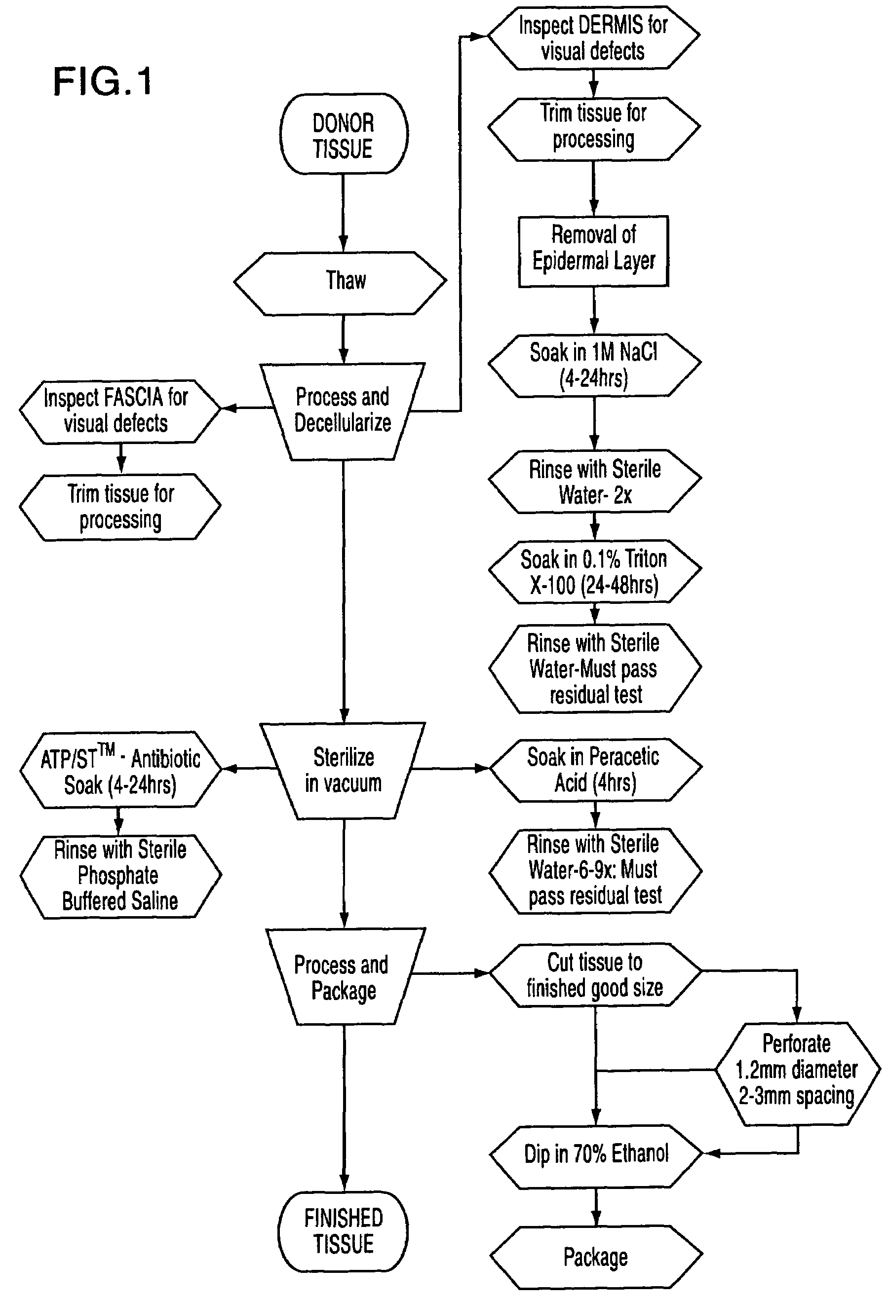
The present invention is a process for preparing soft tissue for implant in a human. The process involves isolating a desired soft tissue sample, decellularizing the tissue, sterilizing the tissue, and packaging it for storage and later use. The invention provides a flexible and decellularized sterilized allograft soft tissue that can be used as an implant by a surgeon. The technical effects of the invention include improved soft tissue preservation and flexibility, as well as reduced risk of immune rejection and improved tissue regeneration.

Problems solved by technology

However, problems exist when there is a transfer of biological material from one individual to another.
Tissue rejection is a significant risk associated with transplantation, even with a good histocompatability match.
These immunosuppressive drugs however, have a narrow therapeutic window between adequate immunosuppression and toxicity.
Prolonged immunosuppression can weaken the immune system, which can lead to a threat of infection.
It can thus be seen that the prior art processes require extensive chemical treatment with a multitude of process steps in an attempt to obtain an acellular soft tissue specimen which has limited shelf life.

Method used


Image

  • Soft tissue processing
  • Soft tissue processing
  • Soft tissue processing

Examples


example 1

Treatment of Fascia

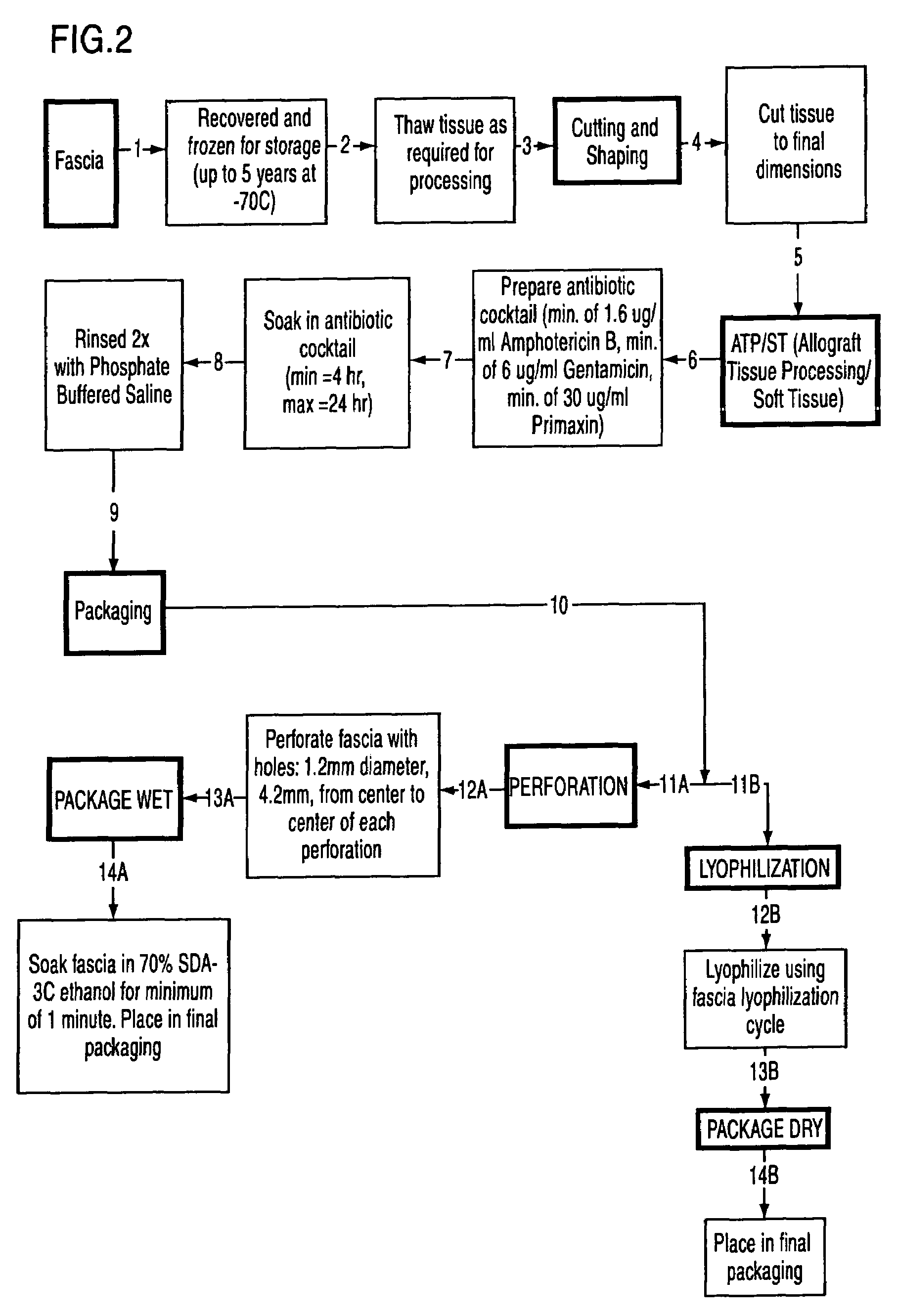
[0033]A desired frozen soft tissue sample is isolated from a suitable donor and then thawed.

[0034]The thawed tissue is processed and decellularized and is inspected for visual defects and trimmed.

[0035]The trimmed tissue sample is sterilized while soaking the tissue in an antibiotic soak for 1.5 to 24 hours and is rinsed with phosphate buffered saline. If desired at the time of sterilization one or more of the following protease inhibitors may be added; Aminoethylbenzenesulfonyl fluoride HCL (Serine Proteases), Aprotinin (broad spectrum, serine proteases), Protease Inhibitor E-64 (Cysteine Proteases), Leupeptin, Hemisulfate Cysteine Proteases and trypsin-like proteases, Pepstatin A (Aspartic Proteases) and Marmistat (MMP2). If desired a solution with pH of 8.0 can be added which will inhibit lysozomal enzymes

[0036]The processed soft tissue is placed into a stainless steel container which is filled with an antibiotic solution. The antibiotic solution is a packet of...

example 2

Treatment of Dermis

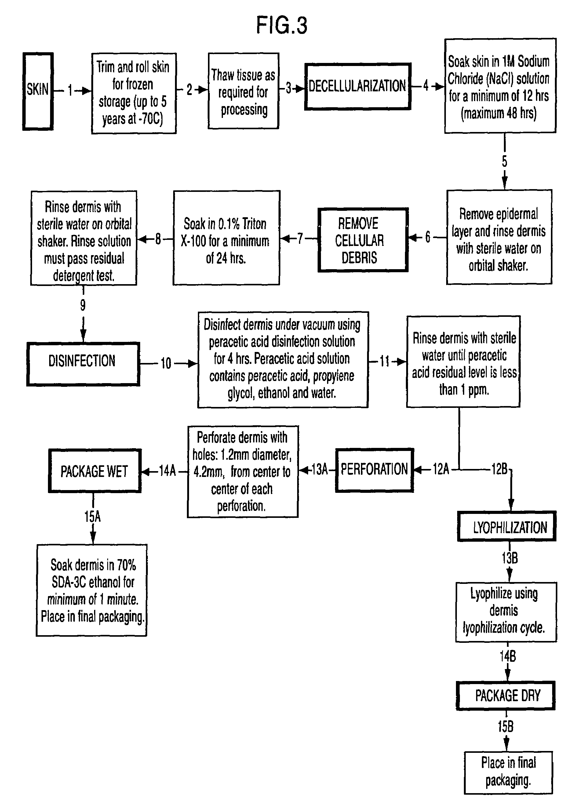
[0040]Frozen donor tissue is then thawed and then rinsed to maintain moisture. The thawed tissue is processed and decellularized. If desired at the time of decellularization one or more of the following protease inhibitors may be added; Aminoethylbenzenesulfonyl fluoride HCL (serine proteases) (25-100 μm, Aprotinin (broad spectrum, serine proteases) (7.5-30 μm), Protease Inhibitor E-64 (cysteine proteases) (0.05-0.0.20 μm), Leupeptin, Hemisulfate (cysteine proteases) (0.05-0.0.20 μm), EDTA, Disodium (0.025-0.0.10 μm), and trypsin-like proteases, Pepstatin A (Aspartic Proteases). Marmistat (MMP2). The thawed tissue is processed and decellularized and is inspected for visual defects and trimmed.

[0041]Once all blood and lipids are removed from the skin, the water is changed with clean sterile water. Impurities are removed from each piece of skin with a scalpel (epidermal side up during this process). Place each skin piece with the epidermal side up on the cutting boa...


PUM

PropertyMeasurementUnit
thickaaaaaaaaaa
diameteraaaaaaaaaa
diameteraaaaaaaaaa

Abstract

The present invention is a process for preparing soft tissue such as tendons, ligaments, cartilage, fascia, dermis, human valves and human veins for implant in a human and removes cellular components and forms an decellular matrix having as major components collagens and elastins while sterilizing the tissue. The process comprises the following steps:(1) isolating from a suitable donor a desired soft tissue sample of the biological material;(2) processing and decellularizing the soft tissue including inspection for visual defects, trimming and soaking the tissue in a detergent depending on whether the tissue is fascia or dermis and rinsing same with sterile water;(3) sterilizing the soft tissue in a vacuum and soaking the tissue in an antibiotic composition or peracetic acid depending on whether the soft tissue is fascia or dermis and rinsing same;(4) processing the tissue by cutting the tissue to size and perforating the tissue; and(5) dipping the tissue in 70% ethanol and packaging the tissue.

Description

RELATED APPLICATIONS[0001]The present application claims priority from U.S. Provisional Patent Application No. 60 / 662,078, filed Mar. 16, 2005.FIELD OF INVENTION[0002]The present invention is generally directed toward methods of treatment of allograft soft tissue including decellularizing and sterilization for implantation into another human being.BACKGROUND OF THE INVENTION[0003]Techniques for restoring structure and function to damaged tissue are used routinely in the area of reconstructive surgery. Tissue transplantation is another way of restoring function by replacing or rebuilding the damaged tissue. However, problems exist when there is a transfer of biological material from one individual to another. Tissue rejection is a significant risk associated with transplantation, even with a good histocompatability match. Immunosuppressive drugs such as cyclosporin and FK506 are usually given to the patient to prevent rejection. These immunosuppressive drugs however, have a narrow th...

Claims


Application Information

Patent Timeline
Patent Type & Authority Patents(United States)
IPC IPC(8): C12N5/00A61K35/32A61K35/35
CPCA01N1/02A01N1/0215A61K35/35A61K35/32A61L27/3687A61L2430/40A61L27/362
Inventor TRUNCALE, KATHERINE GOMESCARTMELL, JEFFREY STUARTSYRING, CARINAVON VERSEN, RUDIGERNGO, MANH-DAN
Owner MUSCULOSKELETAL TRANSPLANT FOUND INC
Learn More