Cytological analysis by raman spectroscopic imaging
a cytological analysis and raman technology, applied in the field of mammalian cellular evaluation, can solve the problems of inability to detect other disease states (including many physiological states) by ordinary cytological methods, inability to perform vivo cytological methods, and inability to achieve the effect of reducing the risk of cancer
- Summary
- Abstract
- Description
- Claims
- Application Information
AI Technical Summary
Benefits of technology
Problems solved by technology
Method used
Image
Examples
example 1
[0152]Raman Scattering Analysis of Bladder Cancer Cells.
[0153]Raman molecular imaging (RMI) was used to distinguish cancerous and non-cancerous bladder cancer cells to demonstrate that RMI is useful for detection of bladder cancer.
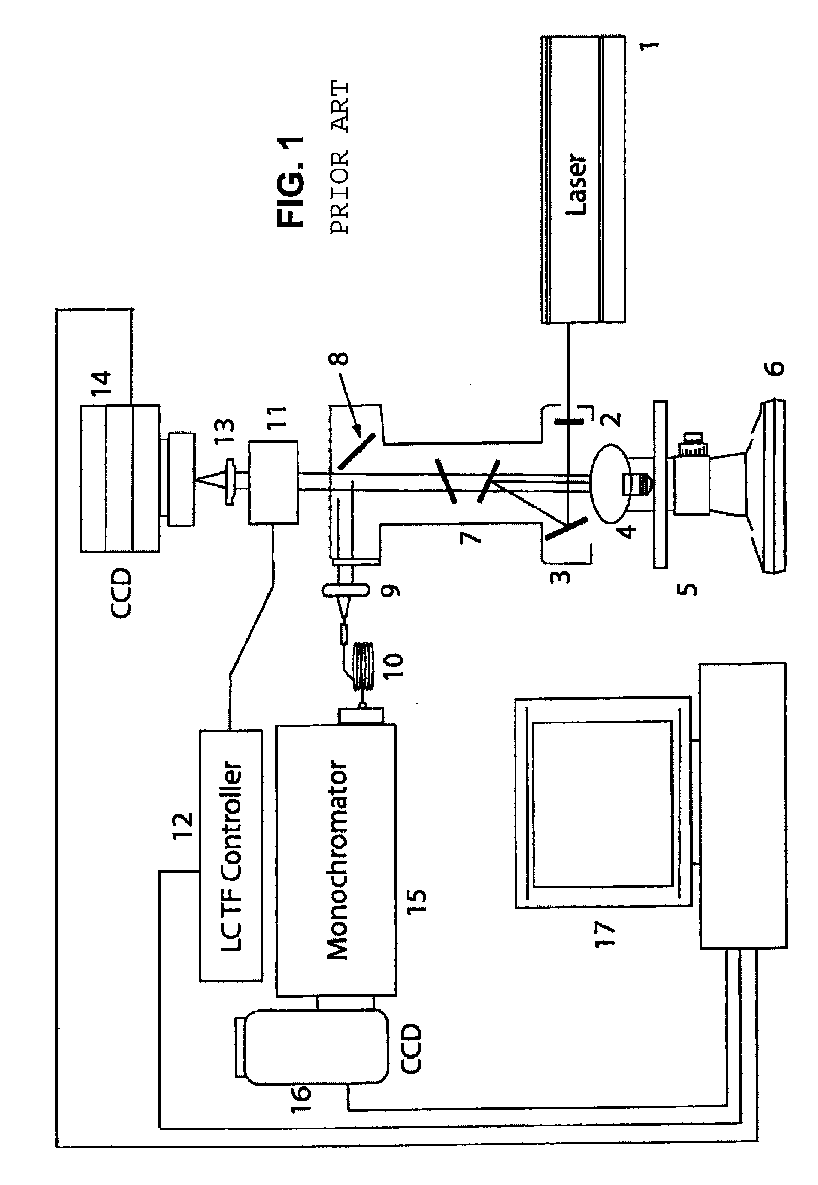
[0154]RMI is an innovative technology that combines the molecular chemical analysis capacity of Raman spectroscopy with the power of high definition digital image microscopic visualization. This platform enables physicians and their assistants to identify both the physical architecture and molecular environment of cells in a urine sample and can complement or be used in place of current histopathological methods.
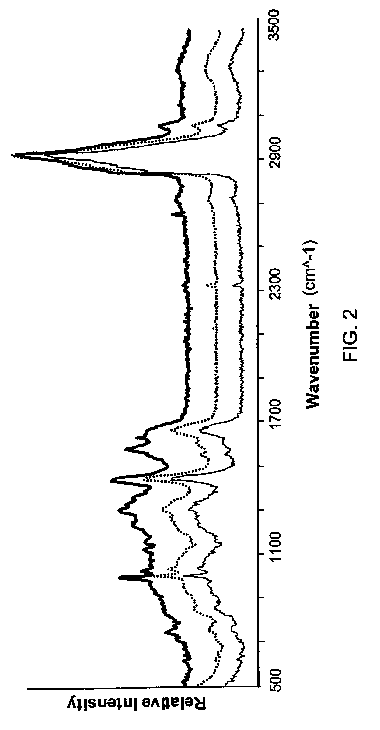
[0155]The data presented in this example demonstrate that the Raman scattering signal from bladder cancer tissue and cells voided in the urine can be identified and be distinguished from normal bladder tissue and cells. Detectable differences between high and low grade tumor cells were observed. These data establish that RMI signatures of bladder c...
example 2
[0167]Raman Scattering Analysis of Red Blood Cells.
[0168]Raman molecular imaging (RMI) was used to distinguish normal and sickled human red blood cells (RBCs).
[0169]Individual RBCs were obtained from two patients, one of whom was known to be afflicted with sickle cell disease (i.e., homozygous for the sickle cell trait gene) and the other of whom was known not to harbor an allele of the gene for the sickle cell trait. Prior to analysis, RBCs were treated by smearing onto an aluminum-coated glass slide and air dried.
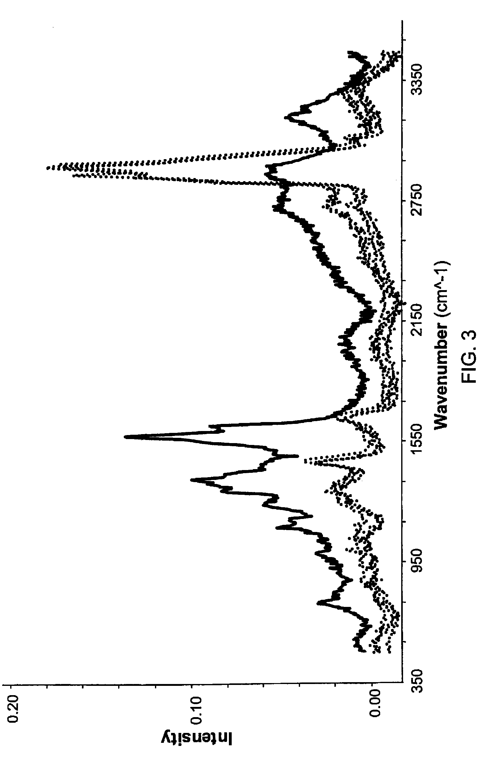
[0170]For each RBC, a visual microscopic determination was made of whether the cell was normal (i.e., normally-shaped) or sickled (i.e., sickle-shaped) using a FALCON (TM) Raman imaging microscope obtained from ChemImage Corp. (Pittsburgh, Pa.). A single Raman spectrum was obtained from a field of view that included 3-5 RBCs using the Raman scattering channel of the FALCON instrument. For samples of sickled RBCs, each field included at least one RBC that exhibited the cre...
example 3
[0173]Raman Scattering Analysis of Cardiac Tissue.
[0174]Raman molecular imaging (RMI) was used to assess cardiac muscle tissue and connective tissue in cardiac tissue samples obtained from patients afflicted with either idiopathic heart failure or ischemic heart failure.
[0175]Human cardiac tissue samples were obtained from five patients afflicted with ischemic heart failure and from five other patients afflicted with idiopathic heart failure. The tissue samples were obtained in the form of small tissue fragments fractured from explanted hearts which were frozen immediately after removal. Approximately 5 millimeter square tissue fragments were embedded in OCT and sliced into 5-10 micron sections. Tissue slices were placed on an aluminum coated slide. Excess OCT was removed with distilled water. Samples were air-dried and evaluated using a FALCON (TM, ChemImage Inc., Pittsburg, Pa.) Raman microscope.
[0176]Each tissue sample was sighted by visible light microscopy a Raman spectrum was ...
PUM
 Abstract
Description
Claims
Application Information
 - R&D
- Intellectual Property
- Life Sciences
- Materials
- Tech Scout
- Unparalleled Data Quality
- Higher Quality Content
- 60% Fewer Hallucinations
Browse by: Latest US Patents, China's latest patents, Technical Efficacy Thesaurus, Application Domain, Technology Topic, Popular Technical Reports.
© 2025 PatSnap. All rights reserved.Legal|Privacy policy|Modern Slavery Act Transparency Statement|Sitemap|About US| Contact US: help@patsnap.com


