
Rapid peptidoglycan-based assay for detection of bacterial contamination of platelets

a platelet contamination and peptidoglycan technology, applied in the field of colorimetric assays and methods, can solve the problems of inability to apply directly for sample testing, severe illness, death, etc., and achieve the effect of rapid detection, sensitive and specific assays, and easy detection of bacterial populations

Active Publication Date: 2009-10-06
IMMUNETICS
View PDF31 Cites 15 Cited by
  • Summary
  • Abstract
  • Description
  • Claims
  • Application Information

AI Technical Summary

Benefits of technology

[0008]To address this need, with specific application to ensuring the safety of platelets for transfusion, we have developed a rapid, sensitive, and specific assay for the detection of bacteria in platelet units. The assay is based on the detection of peptidoglycan, a cell wall component of all bacteria. Present in both Gram-negative and Gram-positive bacteria, peptidoglycan can be used to detect bacterial species known to be frequent contaminants of platelet units as well as less common contaminants or slow growing bacterial pathogens. Further, since peptidoglycan is a major structural component of the cell wall it can be easily and rapidly detected in bacterial populations. The assay may also be used to detect β-glucan, a cell wall component of true fungi, such as yeast and molds.
[0009]In one aspect, the present invention features a rapid, easy, and sensitive method for detecting peptidoglycan (PG) in a sample using a prophenoloxidase cascade system (POC) that forms a red-colored reaction product in the presence of PG. The presence of the red-colored reaction product may be determined rapidly, such as in less than one hour, and easily, such as by visual inspection or spectrophotometrically. In exemplary embodiments, the red-colored reaction product is generated when quinones produced from oxidized substrates of phenoloxidase (PO) react with 3-methyl-2-benzothiozolinone hydrazone (MBTH) or a derivative thereof. In a further embodiment, the detection of PG in platelets may be enhanced by alkaline extraction at an elevated temperature with subsequent neutralization. In a further embodiment, alkaline extraction is used as an efficient approach for the inactivation of various inhibitory factors, which may be present in the plasma or plasma fraction of platelet preparations. In a still further embodiment, alkaline extraction is used for the solubilization of platelet cells, which permits large amounts of platelets in samples to be tested, thus increasing the assay sensitivity.
[0017]In another aspect, the present invention features a kit for the rapid, easy, and sensitive detection of peptidoglycan in a sample. A kit for detecting peptidoglycan in a sample may comprise a prophenoloxidase cascade system, a phenoloxidase substrate that generates a quinone reaction product, and 3-methyl-2-benzothiazolinone hydrazone or derivative thereof. The prophenoloxidase cascade system is obtained from insect plasma or hemolymph, and in exemplary embodiments, is obtained from silkworm larvae plasma. The prophenoloxidase cascade system used in the kit comprises prophenoloxidase activating enzyme, prophenoloxidase, and a serine proteinase cascade. The kit may further comprise a peptidoglycan binding protein. The phenoloxidase substrate that generates a quinone reaction product provided in the kit may be L-3,4-dihydroxyphenylalanine (DOPA), dopamine, or another mono- or dihydroxy-phenolic compound. The kit may further comprise a peptidoglycan standard, wherein the peptidoglycan standard is isolated bacterial peptidoglycan, whole bacterial extract, or inactivated whole bacteria. Still further, the kit may comprise an extraction solution, such as an alkaline extraction solution. The kit may also comprise a neutralization buffer with or without MBTH or derivative thereof dissolved in a neutralization buffer. Where the kit comprises a neutralization buffer without MBTH or derivative thereof dissolved therein. The kit may further comprise a dry detection reagent containing MBTH or a derivative thereof co-lyophilized with a prophenoloxidase cascade system and a phenoloxidase substrate that generates a quinone reaction product. The kit may further comprise a stop reagent, wherein the stop reagent is an acid reagent (e.g., tricholoracetic acid, perchloric acid, or tungstosilicic acid), an inhibitor of phenoloxidase (e.g., phenyl-thiourea), or a strong anionic detergent in sodium or lithium salt forms (dodecylsulphate or laurylsarcosine) alone or in combination with a phenoloxidase specific inhibitor. The kit may still further comprise instructions for spectrophotometric detection or a color-coded scale for visual evaluation as well as a sterile sample tube for performing the reaction.
[0018]In another aspect, the present invention features a kit for the rapid, easy, and sensitive detection of β-glucan in a sample. A kit for detecting β-glucan in a sample may comprise a prophenoloxidase cascade system, a phenoloxidase substrate that generates a quinone reaction product, and 3-methyl-2-benzothiazolinone hydrazone or derivative thereof. The prophenoloxidase cascade system is obtained from insect plasma or hemolymph, and in exemplary embodiments, is obtained from silkworm larvae plasma. The prophenoloxidase cascade system used in the kit comprises prophenoloxidase activating enzyme, prophenoloxidase, and a serine proteinase cascade. The kit may further comprise a β-glucan binding protein. The phenoloxidase substrate that generates a quinone reaction product provided in the kit may be L-3,4-dihydroxyphenylalanine (DOPA), dopamine, or another mono- or dihydroxy-phenolic compounds. The kit may further comprise a β-glucan standard, wherein the β-glucan standard is isolated fungal β-glucan, whole fungal extract, or inactivated whole fungus. Still further, the kit may comprise an extraction solution, such as an alkaline extraction solution. The kit may also comprise a neutralization buffer with or without MBTH or a derivative thereof dissolved in a neutralization buffer. The kit may further comprise a dry detection reagent containing MBTH or derivative thereof co-lyophilized with a prophenoloxidase cascade system and a phenoloxidase substrate that generates a quinone reaction product. The kit may further comprise a stop reagent, wherein the stop reagent is an acid reagent (e.g., tricholoracetic acid, perchloric acid, or tungstosilicic acid), an inhibitor of phenoloxidase (e.g., phenyl-thiourea) or a strong anionic detergent (e.g., dodecylsulfate) alone or in combination with a phenoloxidase specific inhibitor. The kit may still further comprise instructions for spectrophotometric detection or a color-coded scale for visual evaluation as well as a sterile sample tube for performing the reaction.

Problems solved by technology

Even at early time points in the mandatory maximum five-day storage time limit post-collection, microbial growth may reach significant levels.
Bacterial and fungal contamination of many clinical, agricultural or environmental products may lead to severe illness, and even death, if contacted by a subject or administered to a subject.
One limitation of these methods is that they cannot be applied directly for testing of samples where the spectrum of bacterial pathogens is unknown.
Current tests for bacterial and / or fungal contamination involve complicated tasks requiring particular reaction conditions and often take days to complete.
Another barrier to widespread acceptance of various tests for bacterial or fungal contamination is low sensitivity, low specificity, and high cost.
This process can often take up to 72 hours.

Method used


Image

  • Rapid peptidoglycan-based assay for detection of bacterial contamination of platelets
  • Rapid peptidoglycan-based assay for detection of bacterial contamination of platelets
  • Rapid peptidoglycan-based assay for detection of bacterial contamination of platelets

Examples


example 1

Bacteria Spiked Platelets

[0101]In order to assess the sensitivity of the assay, spiking studies were performed with the following bacteria species: Proteus vulgaris, Yersinia enterocolitica, Serratia marcescens (S. marcescens), Enterobacter cloacae, Staphylococcus epidermidis (S. epidermidis), Staphylococcus aureus (S. aureus), Klebsiella pneumoniae, Bacillus cereus, Escherichia coli (E. coli), Proteus mirabilis, Pseudomonas aeruginosa (P. aeruginosa); and Salmonella cholerae. All of these species represent common skin flora. Bacteria species were obtained from the American Type Culture Collection.

[0102]Bacteria were cultured and quantified by reconstituting the bacteria according to the vendor's instructions and grown in Trypticase Soy Broth (Becton Dickinson) overnight at 37° C. Bacteria were washed twice by centrifugation in sterile phosphate buffered saline (PBS) and resuspended in 5 ml sterile PBS. Serial 10-fold dilutions were prepared in PBS by adding 100 μl bacterial suspens...

example 2

Time Course Study of Bacterial Growth in Platelets

[0108]In a different approach to assessment of assay sensitivity, a time course study of bacterial growth in platelets was performed using Assay Method 2. In this series of experiments, assay results vs. CFU / ml were determined at increasing times following inoculation of a platelet bag with bacteria. Platelets from two units were removed from platelet bags and placed in sterile 50 ml conical tubes for ease of sampling over time. At time zero, one unit was spiked with 10 CFU / ml of S. aureus (indicated as SA in FIG. 8), and the other was spiked at 10 CFU / ml of P. aeruginosa (indicated as PA in FIG. 8). Immediately after spiking, one ml samples were taken from each. Following incubation for 20 hours and 48 hours, one ml samples were taken from each. Immediately upon harvest, samples were placed at 4° C. to inhibit further bacterial growth until the time of assay. All samples were processed in parallel as follows: one ml of each was cent...

example 3

Specificity of the Assay for Peptidoglycan

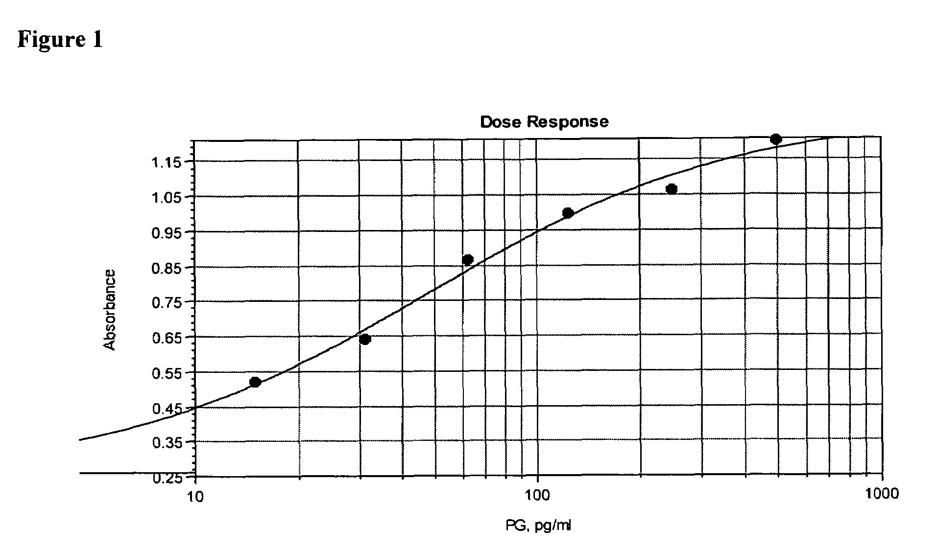
[0110]In order to demonstrate the specificity of the assay for peptidoglycan, a dose response curve was constructed by serially diluting purified peptidoglycan at concentrations from 10 ng / ml to 150 pg / ml into extracted and neutralized platelets as described in Assay Method 1. The absorbance at 490 nm, corrected for background read at 650 nm, is proportional to the peptidoglycan concentration (FIG. 10). A lower limit of 156 pg / ml peptidoglycan was visually detectable.


PUM

PropertyMeasurementUnit
concentrationsaaaaaaaaaa
wavelengthsaaaaaaaaaa
timeaaaaaaaaaa

Abstract

The invention relates to a colorimetric method for detecting bacterial or fungal pathogens by detecting peptidoglycan or (1-3)-β-D-glucan in a sample.

Description

RELATED APPLICATION[0001]This application is a continuation-in-part of International Application No. PCT / US2004 / 035160, filed Oct. 22, 2004, which claims the benefit of U.S. Provisional Application Ser. No. 60 / 516,576, filed Oct. 31, 2003, the content of both of which is hereby incorporated by reference in their entirety.GOVERNMENT SUPPORT[0002]This invention was made with government support under grant No. HL65877 awarded by the National Heart, Lung and Blood Institute. The government may have certain rights in the invention.FIELD OF THE INVENTION[0003]This invention relates to a colorimetric assay and method for rapidly and easily detecting the presence of a bacterial or fungal pathogen in a sample.BACKGROUND OF THE INVENTION[0004]On Mar. 1, 2004, American Association of Blood Banks (AABB) standards mandated that United States blood centers commence testing all platelet units for bacterial contamination. This new standard was based on the significant risks to transfusion patients ...

Claims


Application Information

Patent Timeline
Patent Type & Authority Patents(United States)
IPC IPC(8): C12Q1/26
CPCC12Q1/04C12Q1/26G01N2400/24C12Q2304/20
Inventor KOVALENKO, VICTORLEVIN, ANDREW E.BEAUSANG, LEE ANNE
Owner IMMUNETICS
Learn More