
Dosimeter fitting wear and body surface exposure dose distribution measuring method and apparatus using the same

a technology of exposure dose and wear and body surface, which is applied in the direction of radiation particle tracking, optical radiation measurement, therapy, etc., can solve the problems of increasing the skin dose of patients, affecting treatment activities, and unable to make contact with the patient's skin surface completely with the film, etc., to achieve excellent effects, hinder treatment activities, and easily attached and detached

Active Publication Date: 2009-06-02
NAT INST FOR QUANTUM & RADIOLOGICAL SCI & TECH +1
View PDF7 Cites 18 Cited by
  • Summary
  • Abstract
  • Description
  • Claims
  • Application Information

AI Technical Summary

Benefits of technology

The present invention provides a dosimeter fitting wear and a method for measuring body surface exposure to radiation. The wear is made of a stretchable material that can cover measuring positions on the body and absorb the difference in size of the subject. The wear can be easily attached and detached from the subject and the dosimeters can be housed in sealing bag-like members to prevent contamination. The measured values of the dosimeters can be processed into an exposure dose distribution map and reported to the medical institution. The invention provides a convenient and efficient way to measure body surface exposure to radiation during treatment.

Problems solved by technology

However, due to its longer X-ray fluoroscopy time and increased number of times of photography, the skin dose of a patient increased, and cases of serious skin lesions were reported.
However, with the method (1) in which a film is laid under a patient, it is impossible to make completely closely contact with the patient's skin surface with the film, so that if the patient moves, correct values cannot be obtained.
Furthermore, those measured by this method are only the X-ray doses transmitted through the film laid on the patient bed, so that the obtained values cannot accurately reflect radiation doses at various positions on the three-dimensional body surface of the patient according to X-rays that may be irradiated from various directions of the front, rear, left, and right sides.
On the other hand, the method (2) in which dosimeters are attached onto the surface of the patient's body requires a great deal of work of attaching a number of dosimeters one by one to the surface of the patient's body, and this obstructs actual medical activities, and dose measurement for a number of patients while covering the entire body surfaces in detail involves great difficulty even if it is performed as a clinical study.

Method used


Image

  • Dosimeter fitting wear and body surface exposure dose distribution measuring method and apparatus using the same
  • Dosimeter fitting wear and body surface exposure dose distribution measuring method and apparatus using the same
  • Dosimeter fitting wear and body surface exposure dose distribution measuring method and apparatus using the same

Examples


first embodiment

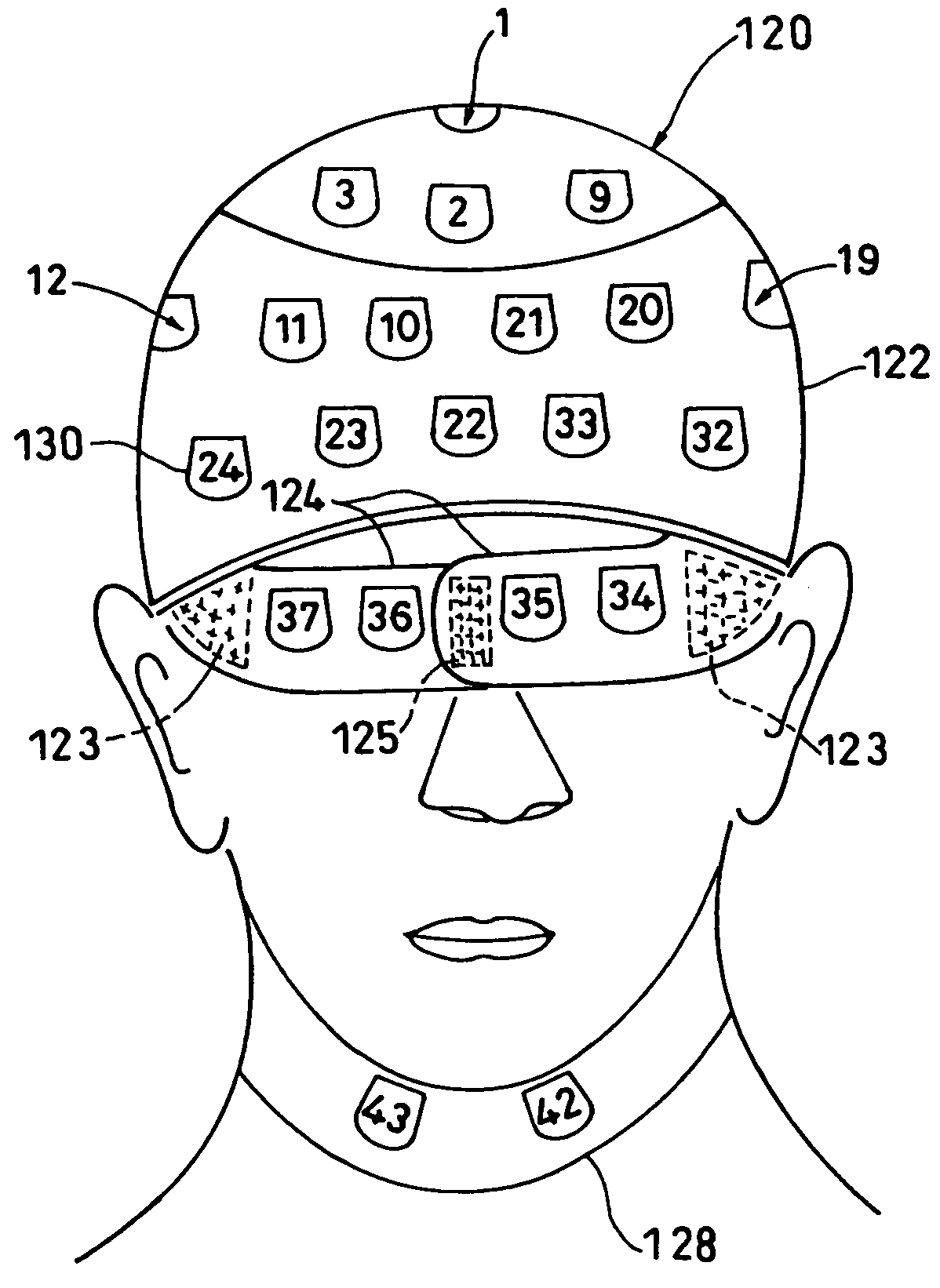
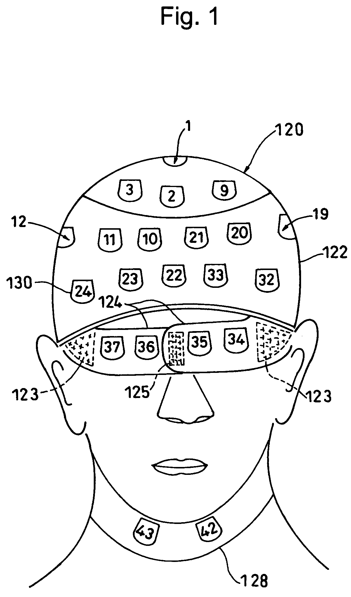
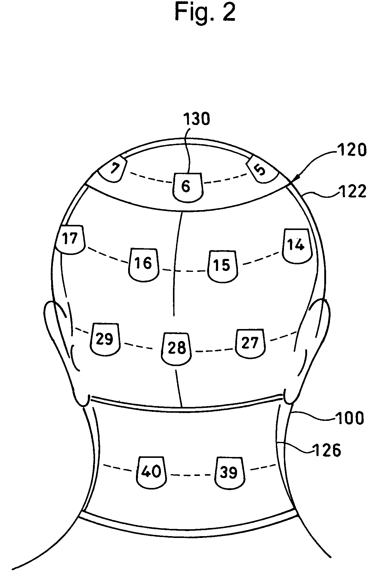
[0060]the present invention is suitable for exposure dose distribution measurement of the head of a patient, and includes, as shown in FIG. 1 (front view showing a fitting state on the head of a patient), FIG. 2 (back view of the same), FIG. 3 (right side view of the same), and FIG. 4 (plan view of the same), a cap-shaped wear main body 120 which covers the head 100 of a patient and can be detachably fitted on the head and a number (forty-three in this embodiment) of dosimeter housing pockets (also referred to as pockets, simply) 130 arranged at positions that come to measuring positions on the outer side of the wear main body 120 when fitted on the head of the patient.

[0061]The wear main body 120 includes a semispherical portion 122 which is in a simple semispherical shape like a swimming cap and covers the head of a patient, an eye-fitting belt 124 which has both ends held on the semispherical portion 122 and is separable at the center, an apron 126 (see FIG. 2 and FIG. 3) hung re...

second embodiment

[0074]Next, the present invention in a T-shirt shape suitable for chest exposure dose distribution measurement will be described in detail.

[0075]The T-shirt-shaped wear main body 150 of this embodiment includes, as shown in FIG. 12 (FIG. 12a front view and FIG. 12b developed view before being fitted on a patient) and FIG. 13 (FIG. 13a front view and FIG. 13b back view showing a fitted state on a patient), a trunk portion 152 which is for covering the chest of the patient and can be wound around the trunk of the body of the patient by being separated at the front chest, arm portions 154 which are integrated with the trunk portion 152 and can be wound around the brachia 110 of the patient by being separated on the inner sides of the brachia, a neck-fitting belt 156 which has both ends held on the trunk portion 152 and can be wound around the cervix of the patient, and dosimeter housing pockets 130 similar to those of the first embodiment provided on the outer sides of the respective p...

third embodiment

[0080]In this embodiment, the trunk portion 152 and the arm portions 154 are integrated, so that reproducibility of the dosimeter positions to be housed in the pockets of the arm portions 154 is high. As in the third embodiment shown in FIG. 14, the vest shape including separated arm portions 154 is also allowed.

[0081]In any of the first through third embodiments, as the wear main body 120 or 150, varied sizes for children or adults can be prepared. Instead of the surface fasteners, resin-made fasteners or tape-like buttons may also be used.

[0082]When performing measurement, as shown in FIG. 15, dosimeter fitting wears 120 and 150 housing dosimeters 140 initialized at the dosimeter service institution 200 in the pockets 130 are delivered to the medical institution 210 from the dosimeter service institution 200, and the dosimeter fitting wears 120 and 150 after being fitted and used on the patient at the medical institution 210 are returned to and recovered by the dosimeter service i...


PUM


Abstract

Dosimeters 140 are housed in dosimeter housing pockets 130 of a dosimeter fitting wear including: wear main bodies 120, 150 which cover measuring positions on at least subject bodies 100, 102, and 110, and can be detachably fitted on the subject bodies, and the dosimeter housing pockets 130 provided at positions that come to the measuring positions when the wear main bodies are fitted on the subject bodies, and then the wear main bodies are fitted on the subject bodies. Thereby, a number of dosimeters can be easily fitted on or detached from the surfaces of subject bodies without hindering medical activities, and set positions of the dosimeters can be easily reproduced.

Description

TECHNICAL FIELD[0001]The present invention relates to a dosimeter fitting wear and body surface exposure dose distribution measuring method and apparatus using the same which realize easy measurement of exposure doses on a body surface for grasping the real condition of medical exposure and preserving it as a medical record in the medical field using a radiolucent finding.BACKGROUND ART[0002]IVR (interventional radiology) is for treating a human body from the inside of a blood vessel without incision of the human body by advancing a catheter inserted into the blood vessel from the femoral or forearm artery to a portion where an aneurysm or stenosis occurs in the brain or heart and placing a coil or a stent there, or advancing the catheter to a cancerous portion and dosing an embolic material or anticancer drug bringing about a high treatment effect with less invasion in comparison with other treatment methods, so that recently, IVR has produced successful results as a treatment meth...

Claims


Application Information

Patent Timeline
Patent Type & Authority Patents(United States)
IPC IPC(8): G01T1/00
CPCA61B5/6805A61N5/1048G01T1/02A61B6/12
Inventor MORITAKE, TAKASHIFUJISAKI, SABUROKOGUCHI, YASUHIROMIZUSHIMA, HIROSHIMATSUNO, KIYOSHIUCHIDA, TAKASHI
Owner NAT INST FOR QUANTUM & RADIOLOGICAL SCI & TECH
Learn More