
System and method for locating and accessing a blood vessel

a blood vessel and imaging system technology, applied in the field of infrared imaging for viewing and accessing subcutaneous blood vessels, can solve the problems of difficult, if not impossible, to locate subcutaneous blood vessels for injection, pain, trauma, etc., and achieve accurate and rapid location, easy to locate, and high quality images.

Inactive Publication Date: 2009-05-12
SYRIS SCI
View PDF3 Cites 65 Cited by
  • Summary
  • Abstract
  • Description
  • Claims
  • Application Information

AI Technical Summary

Benefits of technology

"The present invention is an imaging system for locating subcutaneous blood vessels in a region under the surface of the skin. The system includes an infrared emitter, infrared detector, computing unit, display device, and power source. The infrared emitter illuminates the region with waves of infrared light, and the infrared detector receives the reflected light and outputs a signal. The computing unit processes the signal and outputs the result as enhanced images in real-time. The system also includes a headset with infrared detector, computing unit, display device, and power source. The method involves preparing the body target area, putting on the headset, powering up the system, locating a target blood vessel, and inserting a hypodermic needle into the target blood vessel. The system and method provide a non-invasive and efficient way to locate blood vessels during medical procedures."

Problems solved by technology

Though IV is generally considered routine, there are a number of situations in which inhibited IV access can be painful, traumatic, or even dangerous to patients.
These include conditions in which subcutaneous blood vessels are difficult to locate because of patient characteristics or environmental conditions.
For example, in battlefield conditions, where lighting is limited, it may be difficult, if not impossible, to locate subsurface blood vessels for injection.
Medical practitioners often encounter difficulty in gaining IV access in a significant portion of the patient population for which subsurface blood vessels are obscured.
Difficulties arising in these populations are demonstrated by the numbers: first-stick success rates in children and infants are currently 30%, which indicates that for 70% of the time, IV access in these populations requires more than one stick attempt.
In neonates, more than 90% of IV catheters must be removed prematurely, mainly because of the improper placement of the catheters.
Difficulties with IV access are encountered not only in locating the subsurface blood vessels, but also in complications that arise from improper insertion of needles or catheters in target blood vessels.
With the cost of operating room time approximately $14,000 per hour, delayed IV access can significantly increase the expense of both operating and office-based medical procedures.
A loss of time or inability to obtain IV access can mean the difference between life and death or, at a minimum, cause significant physical and psychological trauma.
Further complicating matters, loss of patient blood and blood pressure in trauma situations can make locating subsurface blood vessels extremely difficult.
In cases where catheters, cannulas, and / or IV drips are used in patient treatment, these devices typically remain in a patient's blood vessel for a long period of time.
Constant relocation of these devices over a long-term hospital stay may result in a need for medical practitioners to access less-optimal blood vessels, after more prominent blood vessels have been used.
Often, these less prominent blood vessels can not easily be found by visual and tactile clues, and accessing them may require multiple sticks to the patient, which thereby causes the patient physical and emotional pain and trauma.
Inhibited IV access can also subject medical practitioners to legal liability risk, by contributing to the complications associated with improper, ineffective, or delayed IV access.
Unfortunately, traditional methods of IV location and access may require years of trial-and-error practice and thereby delay critical healthcare, which increases healthcare costs and possibly jeopardizes patient health.
While the '622 patent describes an IR blood vessel viewer, the '622 patent utilizes an analog signal processor, which is not adequate for supporting the digital algorithms needed for true image enhancement and visualization.
However, because of the need to provide near “real time” images, these devices primarily depend on raw images, or images marginally enhanced by traditional analog means, which are of relatively poor quality for venepuncture accuracy.

Method used


Image

  • System and method for locating and accessing a blood vessel
  • System and method for locating and accessing a blood vessel
  • System and method for locating and accessing a blood vessel

Examples


Embodiment Construction

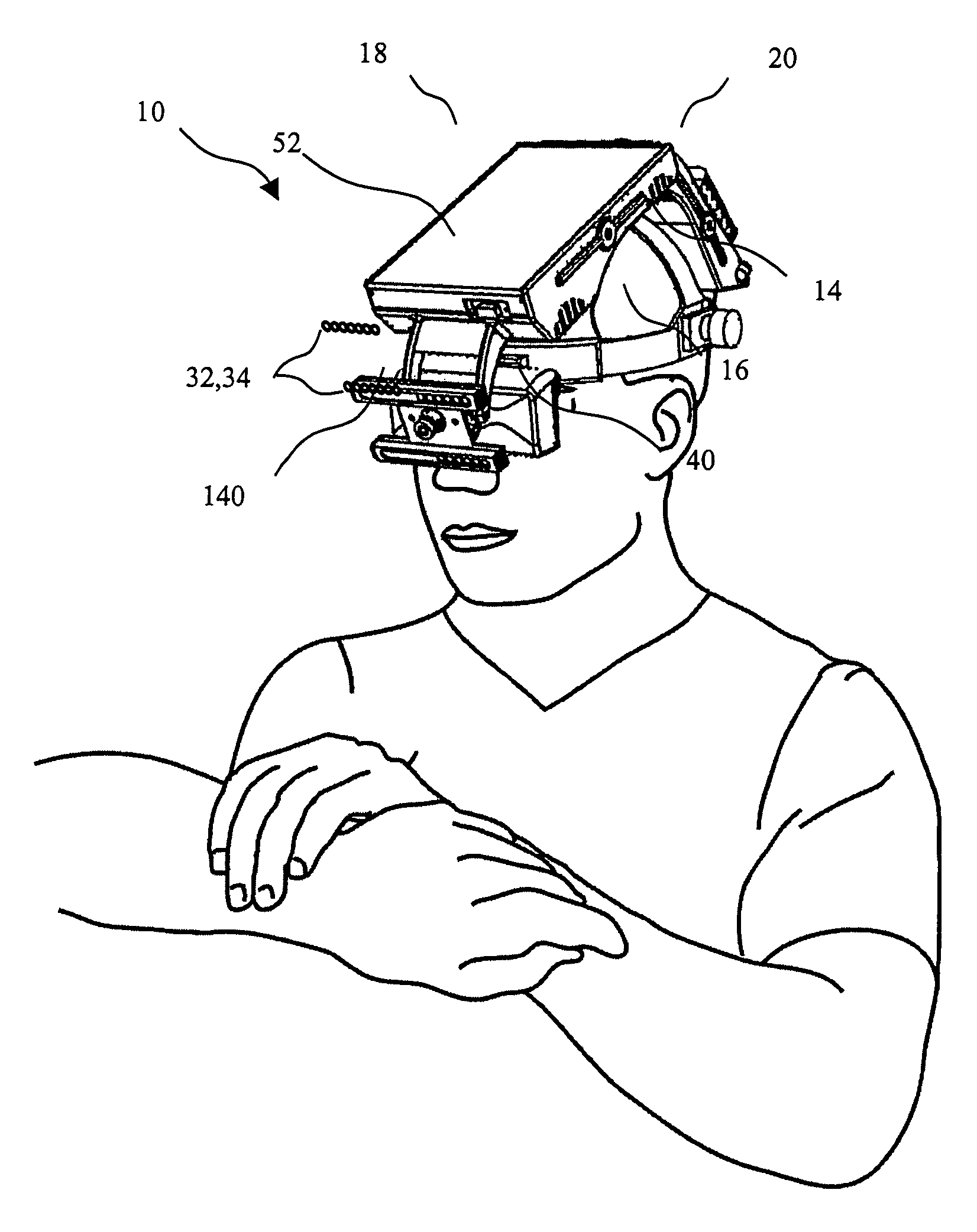
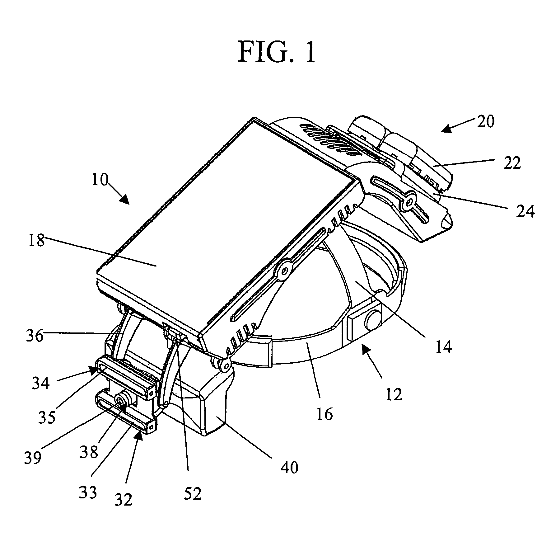
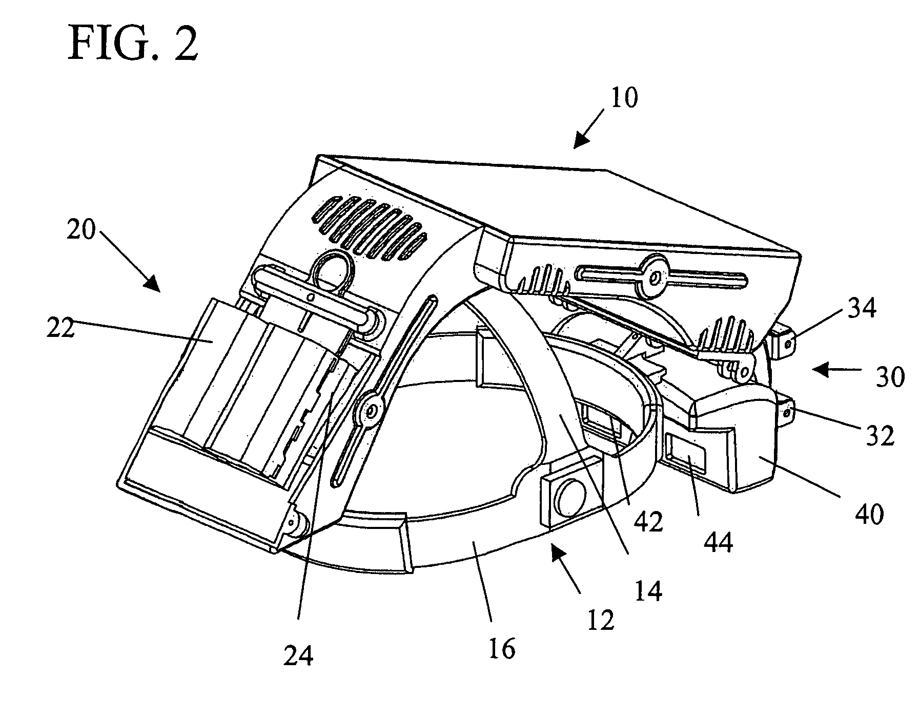
[0044]FIGS. 1-3 show the preferred embodiment of the imaging system 10 of the present invention. The preferred embodiment of the system 10 includes a headset 12 to which all system components are attached. The preferred headset 12 includes two plastic bands 14,16; a vertical band 14 connected to sides of a horizontal band 16. The vertical band 14, holding most of the system components, generally acts as a load-bearing member, while the horizontal band 16 is adjustable such that it snugly fits about the forehead of the person using the system.

[0045]A pivoting housing 18 is attached to the headband 12. The housing 18 is substantially hollow and is sized to house and protect a headset electronics unit 120 disposed therein. Attached to the housing 18 are a power supply 20, an image capture assembly 30, and an enhanced image display unit 40.

[0046]The power supply 20 for the headset electronics unit 120 preferably includes two rechargeable lithium ion batteries 22, which are connected to ...


PUM


Abstract

An imaging system for locating subcutaneous blood vessels and a method for locating subcutaneous blood vessels using the system. The system includes at least one infrared emitter an infrared detector, a computing unit that enhances images and outputs enhanced images in substantially real time, a display device for displaying enhanced images, and a power source. The method includes the steps of preparing a body target area, putting on the headset, powering up the system, locating a target blood vessel, inserting a needle into the target blood vessel, and performing the medical procedure.

Description

CLAIM OF PRIORITY[0001]This application is a Continuation-in-Part of U.S. patent application Ser. No. 10 / 760,051, filed Jan. 16, 2004 now abandoned.FIELD OF THE INVENTION[0002]The present invention relates generally to the non-invasive viewing of surface and subsurface blood vessels by use of an infrared imaging. In particular, the present invention relates to an imaging system for viewing and accessing subcutaneous blood vessels and a method of use thereof.BACKGROUND OF THE INVENTION[0003]Intravenous (IV) access is the single most frequently performed invasive medical procedure in the world today. Though IV is generally considered routine, there are a number of situations in which inhibited IV access can be painful, traumatic, or even dangerous to patients. These include conditions in which subcutaneous blood vessels are difficult to locate because of patient characteristics or environmental conditions. For example, in battlefield conditions, where lighting is limited, it may be di...

Claims


Application Information

Patent Timeline
Patent Type & Authority Patents(United States)
IPC IPC(8): G06K9/00G06K9/40G06T5/00G06T5/10
CPCA61B5/0059A61B5/489A61B5/742G06T5/001G06T5/10G06T2207/30101G06T2207/10048A61B5/15003G06T5/20A61B5/150748G06T5/75G06T5/92G06T5/00
Inventor MARCOTTE, RONALDARSENAULT, MARKPELLETIER, DOMINICHEBOLD, WALTERWANER, MILTONFINK, LOUIS
Owner SYRIS SCI
Learn More