
Solid-phase immobilization of proteins and peptides

a technology of proteins and solid supports, applied in the field of solid-phase immobilization of proteins and peptides, can solve the problems of difficult isolation of compounds that bind specifically to biologically important molecules, difficult immobilization and screening of proteins, and inability to use all proteins, etc., to achieve easy purification, less restrictions, and easy to work.

Active Publication Date: 2006-10-24
BRISTOL MYERS SQUIBB CO
View PDF16 Cites 68 Cited by
  • Summary
  • Abstract
  • Description
  • Claims
  • Application Information

AI Technical Summary

Benefits of technology

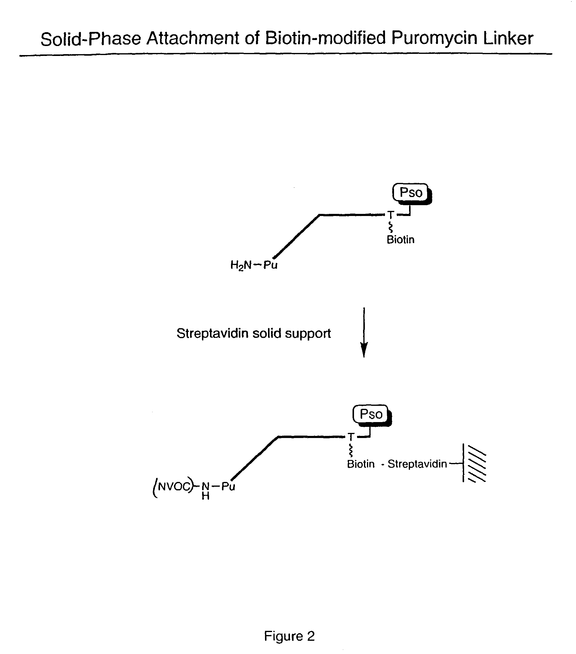
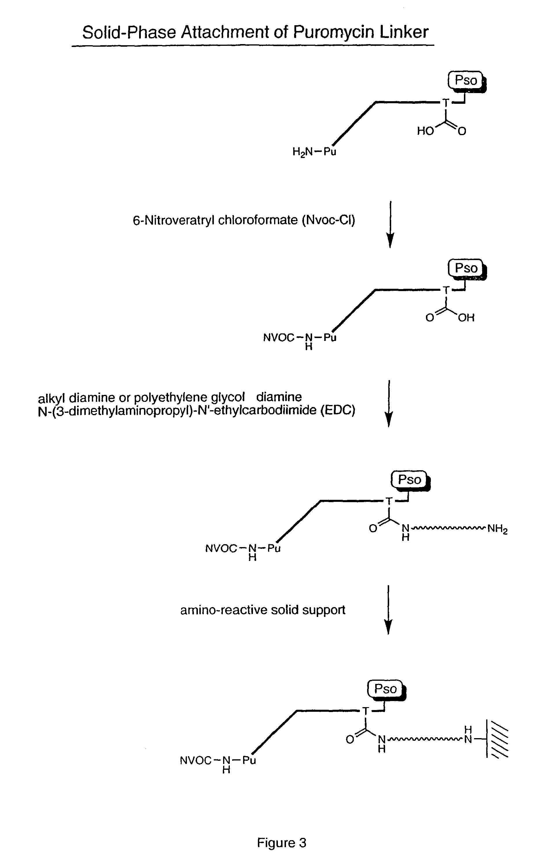
The present invention features a solid support that has a RNA-peptide fusion component immobilized on it, where the peptide is covalently bonded to the RNA through a linker. The RNA-peptide fusion component is attached to the solid support through the linker. The invention also provides a method for screening large collections of compounds to identify potential drug targets. The invention also features a method for synthesizing peptides on a solid support. The technical effects of the invention include improved screening for drug targets and efficient synthesis of peptides.

Problems solved by technology

Unfortunately, the isolation of compounds that bind specifically to biologically important molecules is difficult, in part because the selection of molecules of interest from large ensembles or repertoires is time-consuming, laborious, and costly.
Proteins, however, are more difficult to immobilize and screen.
However, these methods are not possible with all proteins, such as when the protein is insoluble or difficult to purify.
Another problem arises because covalent protein immobilization is usually carried out non-specifically by reaction of functional groups of amino acid residues throughout the protein with appropriately derivatized surfaces.
This approach renders some proteins inactive due to disruption of protein structure and / or inaccessibility of the protein's binding or active sites.
Even if such sites are exposed, the immobilized protein may be too close to the solid support surface to allow for interaction with reagents in the solution phase.
Moreover, even though some of the immobilized proteins do retain functionality, a certain fraction of the protein sample will generally be inactive, resulting in a heterogeneous population.

Method used


Image

  • Solid-phase immobilization of proteins and peptides
  • Solid-phase immobilization of proteins and peptides
  • Solid-phase immobilization of proteins and peptides

Examples


example 1

Periodate Oxidation, Biotinylation of RNA

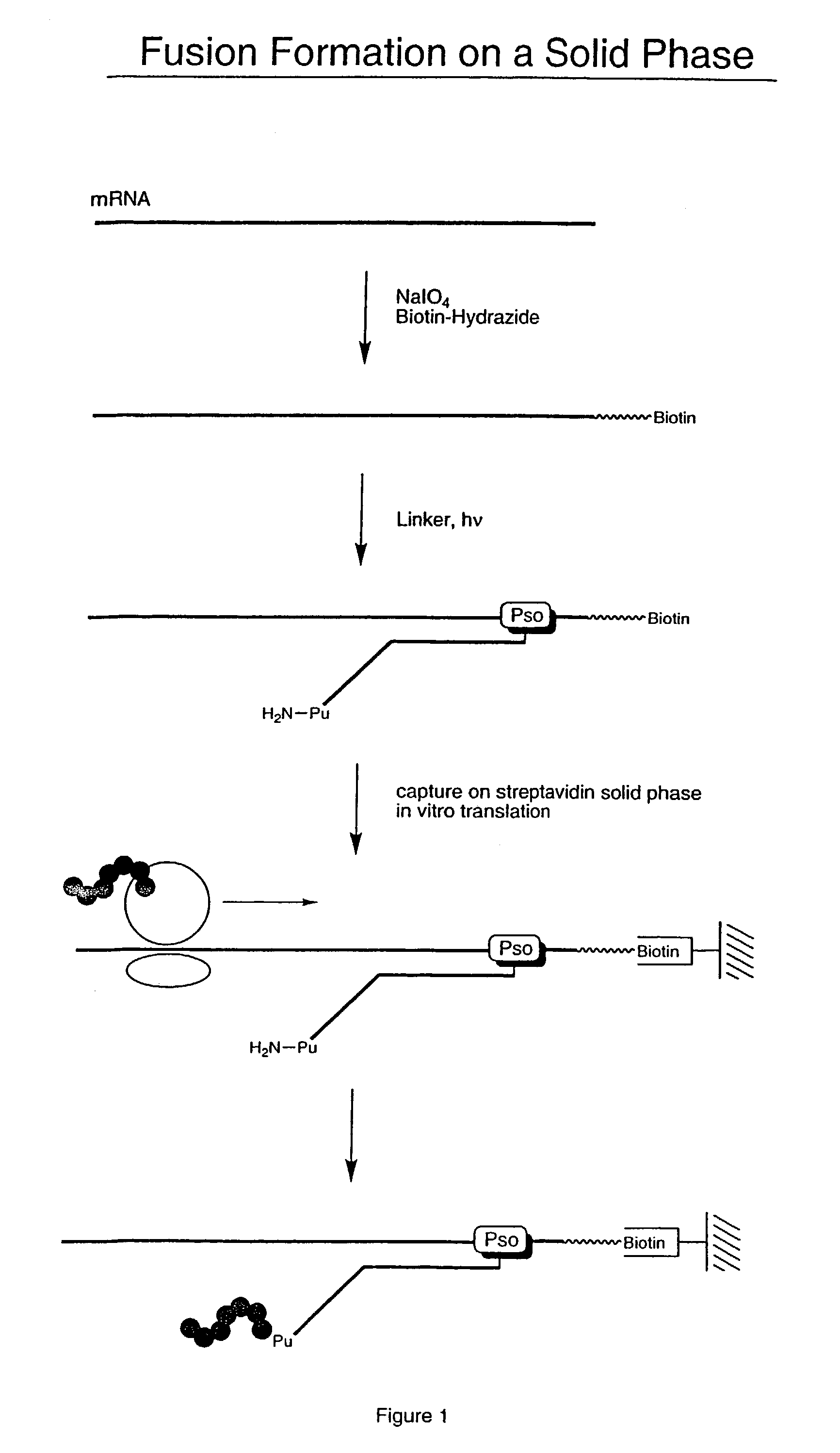
[0058]For this illustrative example, the RNA encodes a peptide having the amino acid sequence:[0059]MVSDVPRDLEVVAATPTSLLISWKTHEVAARYYRITYGETGGNSPVQEFTVPPW ASIATISGLKPGVDYTITVYAVTPLRWTETEAHIPIPINYRT (SEQ. ID NO: 1) The RNA was modified to allow attachment to a solid phase. Modification of the RNA was performed by periodate oxidation of the 3′-terminal ribose (Agrawal Methods in Molecular Biology, Agrawal ed. Vol. 26, Totowa: Human Press, pp. 93–120 (1994)). This was accomplished by mixing 18 μL of RNA (10 μM), 10 μL of NaIO4 (0.5 M), and 3.3 μL of NaOAc (0.1 M) in 68.7 μL of water and incubating for 15 minutes at room temperature.

[0060]Following incubation, the sample was precipitated with a solution of 260 μL H2O, 40 μL NaOAc (3 M), and 1 mL EtOH, and re-suspended in 90 μL NaOAc (0.1 M). 10 μL of Biotin-LC-hydrazide, 50 mM (Pierce, Rockford, Ill.) was added. The mixture was incubated for 2 hours at room temperature and precipitated with NaOAc...

example 2

On-Bead Fusion Formation

[0061]Biotinylated RNA was immobilized on a solid support by mixing 3 μL Bio RNA (and non-biotinylated control), 50 μL buffer (25 mM Tris, pH 7.0; 0.25% Tween), and 25 μL Neutravidin agarose beads. The mixture was incubated at room temperature for 10 minutes and spun in a Microspin column, followed by washing (2×) with 100 μL of buffer (1% BSA in PBS; 0.25% Tween) and washing (2×) with 100 μL of PBS / 0.25% Tween buffer. A translation reaction was performed (see, e.g., Roberts & Szostak, Proc. Natl. Acad. Sci. USA, 1997, 94:12297 or Liu et al., Methods Enzymol., 2000, 318:268) by adding 100 μL lysate mix containing [35S] Methionine. The mixture was rotated for 1 hour at 30° C. The mixture was then spun and 200 μL of 0.5 M KCl and 0.1 M MgCl2 were added to promote RNA-protein fusion formation. The mixture was incubated for 1 hour at room temperature, spun, and washed (5×) with 100 μL PBS buffer. The sample was re-suspended in 100 μL of buffer and the successful ...

example 3

On-Membrane Fusion Formation

[0062]Biotinylated RNA was diluted 1:5, 1:10, 1:20, 1:40, and 1:80 into 25 mM buffer (Tris, pH 7.0; 0.25% Tween-20). 3 μL aliquots were spotted on a dry streptavidin membrane (SAM™ Biotin Capture membrane, Promega, Madison, Wis.). The filter was then washed with buffer (1% BSA in PBS, pH 7.6; 0.25% Tween) and left to dry. A translation reaction was performed by soaking the filter with lysate ([35S] Methionine) and incubating for 1 hour at 30° C. 0.5 M KCl and 0.1 mM MgCl2 were added and the mixture was incubated for 1 hour at room temperature. The filter was then washed for 10 minutes with PBS buffer, rinsed thoroughly, dried, and exposed to a phosphorimager screen for analysis.


PUM

PropertyMeasurementUnit
pHaaaaaaaaaa
insolubleaaaaaaaaaa
covalentaaaaaaaaaa

Abstract

Disclosed herein are methods for immobilizing a peptide or protein on a solid support. The method generally includes the following steps: (a) providing one or more templates attached to a solid support, wherein the one or more templates include (i) an RNA encoding a peptide and (ii) a peptide acceptor-linker linked to the RNA; and (b) subjecting the one or more templates to conditions that support translation and attachment of said peptide to said peptide acceptor, thereby synthesizing the one or more peptides on the solid support. Also disclosed herein are solid supports having at least one RNA-protein fusion component immobilized thereon, methods for generating protein arrays, and methods for screening molecules using these arrays.

Description

CROSS REFERENCE TO RELATED APPLICATIONS[0001]This application claims the benefit of the filing date of U.S. provisional application, U.S. Ser. No. 60 / 333,470, filed Nov. 27, 2001, hereby incorporated by reference.BACKGROUND OF THE INVENTION[0002]In general, the present invention relates to methods for immobilizing peptides and proteins onto a solid support.[0003]Proteins are known to interact specifically with other molecules, such as other proteins, nucleic acids, and small-molecules, based on the three-dimensional shapes and electronic distributions of those molecules. Through these interactions, proteins are responsible for regulating a wide variety of biological processes. The discovery of compounds that interact with proteins can lead to the identification of new drug targets and the development of pharmaceutical products.[0004]One way to search for new drug candidates is to screen large libraries of natural materials or synthesized molecules, using assay techniques that range ...

Claims


Application Information

Patent Timeline
Patent Type & Authority Patents(United States)
IPC IPC(8): C12Q1/68C07H21/02C07H21/04G01N33/53C07K1/04C07K17/06
CPCC07K1/047C07K17/06C12N15/1062
Inventor KURZ, MARKUS
Owner BRISTOL MYERS SQUIBB CO
Learn More