FLT4 (VEGFR-3) as a target for tumor imaging and anti-tumor therapy

Inactive Publication Date: 2006-04-25
VEGENICS PTY LTD
View PDF89 Cites 49 Cited by
  • Summary
  • Abstract
  • Description
  • Claims
  • Application Information

AI Technical Summary

Benefits of technology

[0056]Conjugation of the anti-Flt4 compound to a prodrug that would be targeted to tumor vessels by the anti-Flt4 compound and then activated (e.g., by irradiation) locally at sites of tumor growth also is contemplated. Use of such prodrug strategy has the expected advantage of minimizing side effects of the drug upon healthy lymphatic vessels that express Flt4.
[0060]In a related variation, the invention provides a method for imaging tumors in tissue from a vertebrate organism, comprising the steps of: (a) contacting vertebrate tissue suspected of containing a tumor with a composition comprising an Flt4 binding compound; (b) detecting the Flt4 binding compound bound to cells in said tissue; and (c) imaging solid tumors by identifying blood vessel endothelial cells bound by the Flt4 binding compound, wherein blood vessels expressing Flt4 are correlated with the presence and location of a tumor in the tissue. In one preferred embodiment, the method further comprises steps of contacting the tissue with a second compound (such as an antibody) that specifically binds to a blood vessel endothelial marker (e.g., PAL-E, VEGFR-1, VEGFR-2) that is substantially absent in lymphatic endothelia; and detecting the second compound bound to cells in the tissue; wherein the imaging step comprises identifying blood vessels labeled with both the Flt4 binding compound and the second compound, and wherein blood vessels labeled with both the Flt4 binding compound and the second compound correlate with the presence and location of a tumor in the tissue. It will be appreciated that the use of the second compound helps the practitioner to more rapidly distinguish between blood vessels that are expressing Flt4 and normal lymphatic vessels which express Flt4 on their surface.
[0063]Similarly, the invention includes kits which comprise compounds or compositions of the invention packaged in a manner which facilitates there use to practice methods of the invention. In a simplest embodiment, such a kit includes a compound or composition of the invention packaged in a container such as a sealed bottle or vessel, with a label affixed to the container or included in the package that describes use of the compound or composition to practice the method of the invention. Preferably, the compound or composition is packaged in a unit dosage form. In another embodiment, a kit of the invention includes a Flt4 binding compound packaged together with a second compound that binds to a marker (antigen) that is expressed on the surface of blood vessel endothelial cells but is substantially absent from lymphatic endothelia.

Problems solved by technology

However, lymphatic vessels have been difficult to identify, due to the absence of known specific markers available for them.

Method used


Image

  • FLT4 (VEGFR-3) as a target for tumor imaging and anti-tumor therapy
  • FLT4 (VEGFR-3) as a target for tumor imaging and anti-tumor therapy
  • FLT4 (VEGFR-3) as a target for tumor imaging and anti-tumor therapy

Examples


example 1

Isolation and Characterization of cDNA Clones Encoding Flt4

Materials and Methods

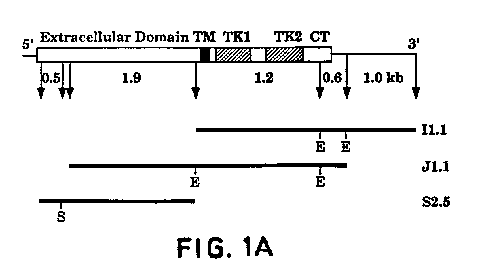
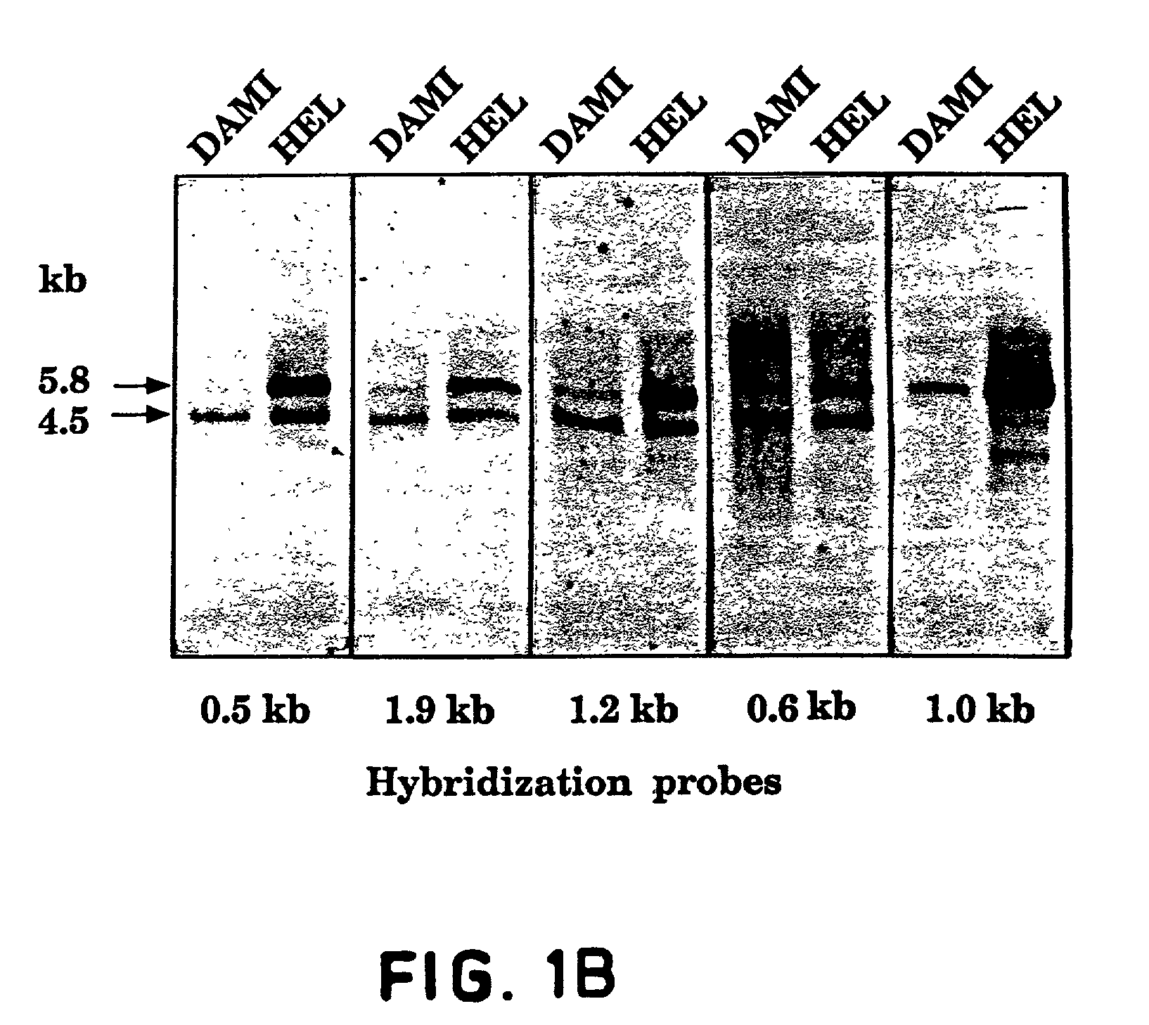
[0149]An oligo-dT primed human HEL cell cDNA library in bacteriophage lambda gt11 [A kind gift from Dr. Mortimer Poncz, Childrens Hospital of Philadelphia, Pa.; Poncz et al., Blood, 69: 219–223 (1987)] was screened with a cDNA fragment PCR-amplified from the same library [Aprelikova et al., Cancer Res., 52: 746–748 (1992)]. Positive plaques were identified and purified as described [Sambrook et al., Molecular Cloning—A Laboratory Manual, Cold Spring Harbor Laboratory Press, (1989)]. cDNA inserts of bacteriophage lambda were isolated as EcoRI fragments and subcloned into a GEM3Zf(+) plasmid (Promega). The entire Flt4 protein coding region was isolated. Three overlapping clones isolated from the HEL-library (as illustrated in FIG. 1) were sequenced using the dideoxy chain termination method with oligonucleotide primers designed according to the sequences obtained. All portions of the cDNAs were sequenced o...

example 2

Preparation of an Anti-Flt4 Antisera

[0155]A 657 base pair EcoRI fragment encoding the predicted C-terminus of Flt4 short form was cloned in-frame with the glutathione-S-transferase coding region in the pGEX-1λT bacterial expression vector (Pharmacia) to produce a GST-Flt4 fusion protein in E. coli. The resulting fusion protein was produced in bacteria and partially purified by glutathione affinity chromatography according to the manufacturer's instructions. This protein was used in immunization of rabbits in order to produce polyclonal antibodies against Flt4. Antisera were used after the third booster immunization.

example 3

Expression of Flt4 in COS Cells

Materials and Methods

[0156]The full-length Flt4 protein coding sequence (combined from three clones, FIG. 1) was inserted into the HindIII-BamHI site of SVpoly mammalian expression vector [Stacey et al., Nucleic Acids Res., 18: 2829 (1990)] construct SV14-2. The expression vectors (SV-FLT4 short and SV-FLT4 long, containing the respective forms of Flt4 cDNA) were introduced into COS cells by DEAE-dextran transfection method [McCutchan et al., J. Natl. Cancer Inst., 41: 351–357 (1968)]. Two days after transfection, the cells were washed with phosphate-buffered saline (PBS) and scraped into immunoprecipitation buffer (10 mM Tris pH 7.5, 50 mM NaCl, 0.5% sodium deoxycholate, 0.5% Nonidet P40, 0.1% SDS, 0.1 TIU / ml Aprotinin). The lysates were sonicated, centrifuged for 15′ at 10,000×g and incubated overnight on ice with 3 ml of the antisera. Protein A sepharose (Pharmacia) was added and the incubation was continued for 30′ with rotation. The precipitates w...


PUM

PropertyMeasurementUnit
Fractionaaaaaaaaaa
Fractionaaaaaaaaaa
Fractionaaaaaaaaaa

Abstract

The present invention provide purified Flt4 receptor tyrosine kinase polypeptides and fragments thereof, polynucleotides encoding such polypeptides, antibodies that specifically bind such polypeptides, and uses therefor.

Description

[0001]This application is a continuation-in-part of U.S. patent application Ser. No. 09 / 169,079 filed Oct. 9, 1998; now U.S. Pat. No. 6,824,777, which is a continuation-in-part of U.S. patent application Ser. No. 08 / 901,710, filed Jul. 28, 1997, now U.S. Pat. No. 6,107,046; which is a continuation-in-part of both U.S. patent application Ser. No. 08 / 340,011, filed Nov. 14, 1994, now U.S. Pat. No. 5,776,755; and U.S. patent application Ser. No. 08 / 257,754, filed Jun. 9, 1994, now abandoned; the latter two of which in turn are continuations-in-part of U.S. patent application Ser. No. 07 / 959,951, filed on Oct. 9, 1992, now abandoned. All of these applications are incorporated herein by reference, in their entireties.FIELD OF THE INVENTION[0002]The present invention relates generally to genes for receptors, specifically genes for receptor tyrosine kinases, their insertion into recombinant DNA vectors, and the production of the resulting proteins in host strains of microorganisms and host...

Claims


Application Information

Patent Timeline
IPC IPC(8): C07K14/00C07H21/04C12N15/00C12N15/12C12N15/63A61K38/00A61K38/17A61K38/19C07K14/52C07K14/71C07K16/28
CPCC07K14/52C07K14/71C07K16/18C07K16/28C07K16/2863C07K16/22A61K2039/505A61K2039/507C07K2319/30C07K2316/96C07K2319/00C07K2317/76
Inventor ALITALO, KARIAPRELIKOVA, OLGAPAJUSOLA, KATRIARMSTRONG, ELINAKORHONEN, JAANAKAIPAINEN, ARJA
Owner VEGENICS PTY LTD
Why Eureka
Try Eureka