
Integrated digital dental x-ray system

a digital x-ray and diagnostic system technology, applied in the field of integrated digital x-ray diagnostic system, can solve the problems of large image sensor arrangement, need of precise amplitude and timing of shifting clocks, and significant technical limitations in the art, so as to improve the productivity and workflow efficiency of the dental office, increase the quality of x-ray images, and efficiently capture, view and manipulate.

Active Publication Date: 2006-02-28
MIDMARK
View PDF20 Cites 80 Cited by
  • Summary
  • Abstract
  • Description
  • Claims
  • Application Information

AI Technical Summary

Benefits of technology

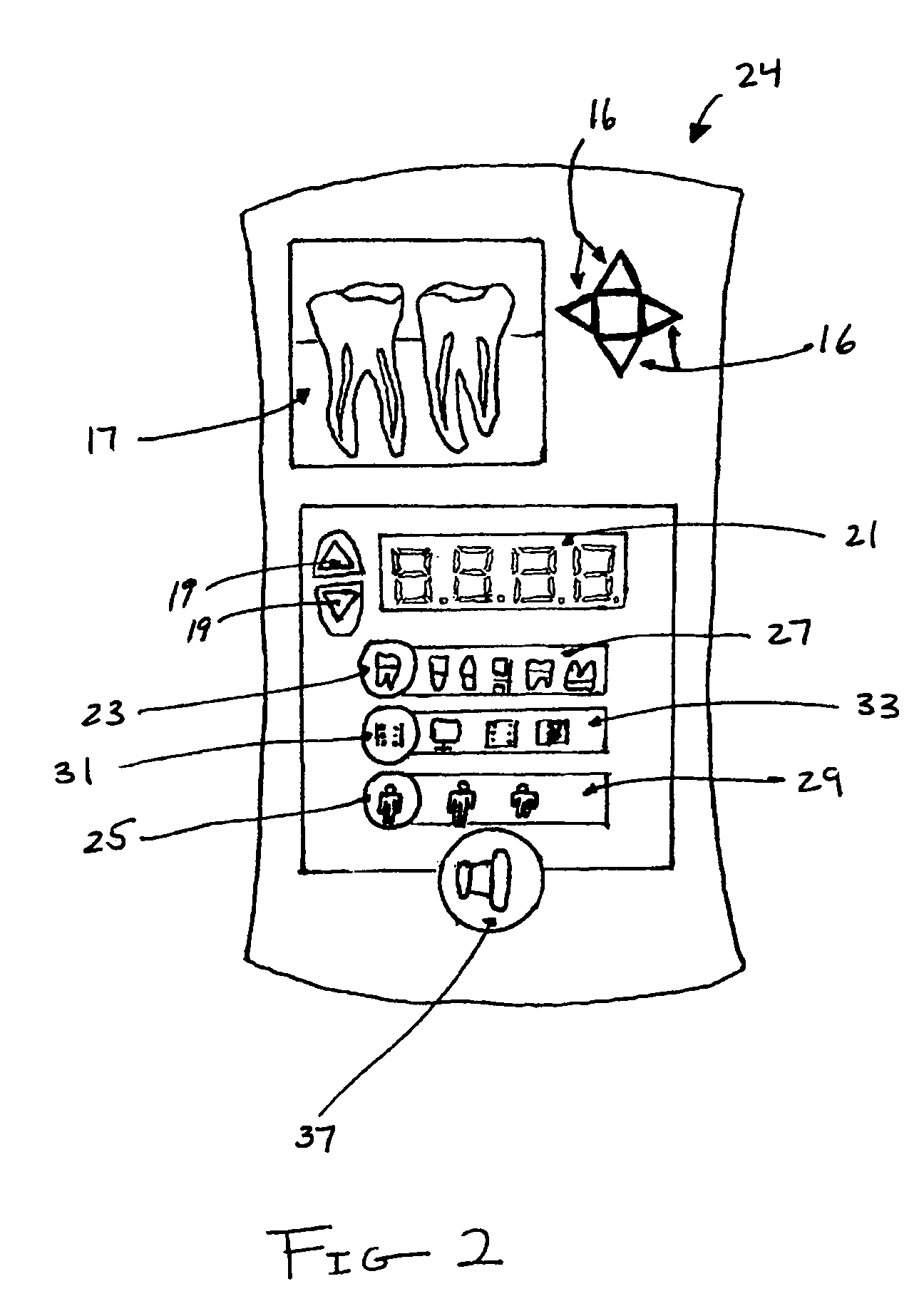
[0012]It is another object of the present invention to provide an integrated digital x-ray system which provides electronic signal transduction of the exposure settings of the x-ray source, thereby eliminating the need for radiation sensors disposed in or about the image detector and substantially increasing the quality of the x-ray images as a result of a reduction of noise accumulating in the detector prior to the activation of the x-ray source.
[0013]It is yet another object of the invention to provide a system and method for taking digital radiographs whereby pre-determined exposure times are provided to the system based on certain characteristics of a patient's anatomy, thereby tailoring the exposure to the particular patient.
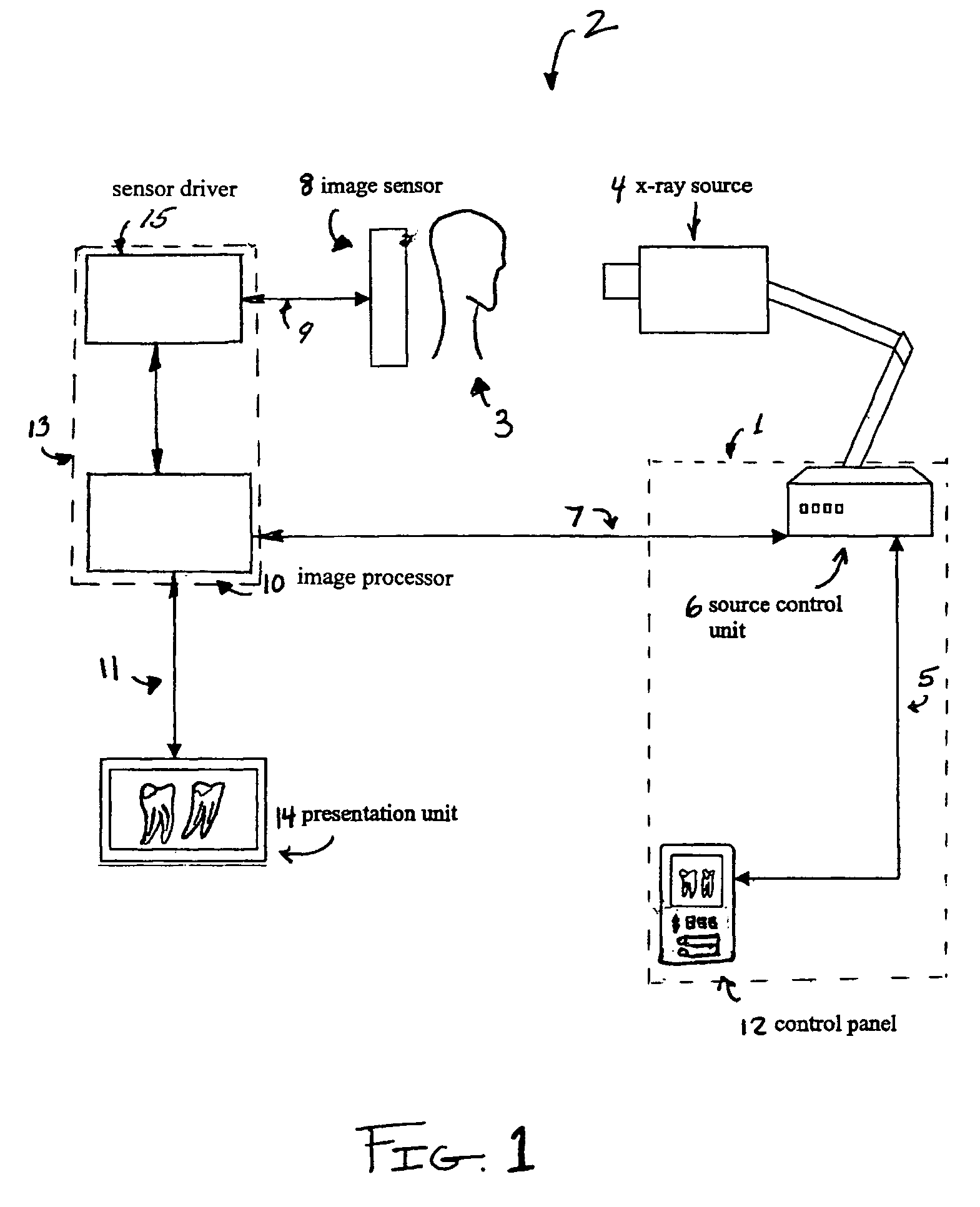
[0014]It is still another object of the invention to provide a system and method of displaying the processed x-ray images in accordance with the invention to allow technicians to efficiently capture, view and manipulate the images without having to leave their post, thereby increasing the productivity and workflow efficiency of the dental office.

Problems solved by technology

It has also, however, led to significant technical limitations in the art.
The use of supplementary sensing elements adds size and complexity to the image sensor arrangement.
The disadvantage of this method is the need of precise amplitude and timing for the shifting clocks in order to avoid loss of signal and image smearing.
Additionally, both embodiments do not utilize customized exposure times calculated based on varying categories of teeth (i.e. incisors, canines, pre-molars and molars), and most importantly, varying anatomies of the patient (adult or child).
This lack of tailored exposure settings may increase the risk of over-exposing children.
The main disadvantages are the lower dynamic range, high pattern noise, lower resolution, high dark current and fair quantum efficiency compared with the CCD image sensors.
The charged CCDs lead to heightened noise accumulation along an unregulated exposure integration cycle, which detrimentally affects the quality of the resultant image.
However, there are obvious drawbacks in having stationary and remote display arrangements, the most evident being the disruption in the efficiency of dental office workflow.
In many instances, however, the first take does not suffice, requiring the technician to repeat the procedure until a usable image is obtained.
This process leads to inefficiencies and decreased productivity in the dental office, as well as increased exposure of patients to x-radiation.

Method used


Image

  • Integrated digital dental x-ray system
  • Integrated digital dental x-ray system
  • Integrated digital dental x-ray system

Examples


Embodiment Construction

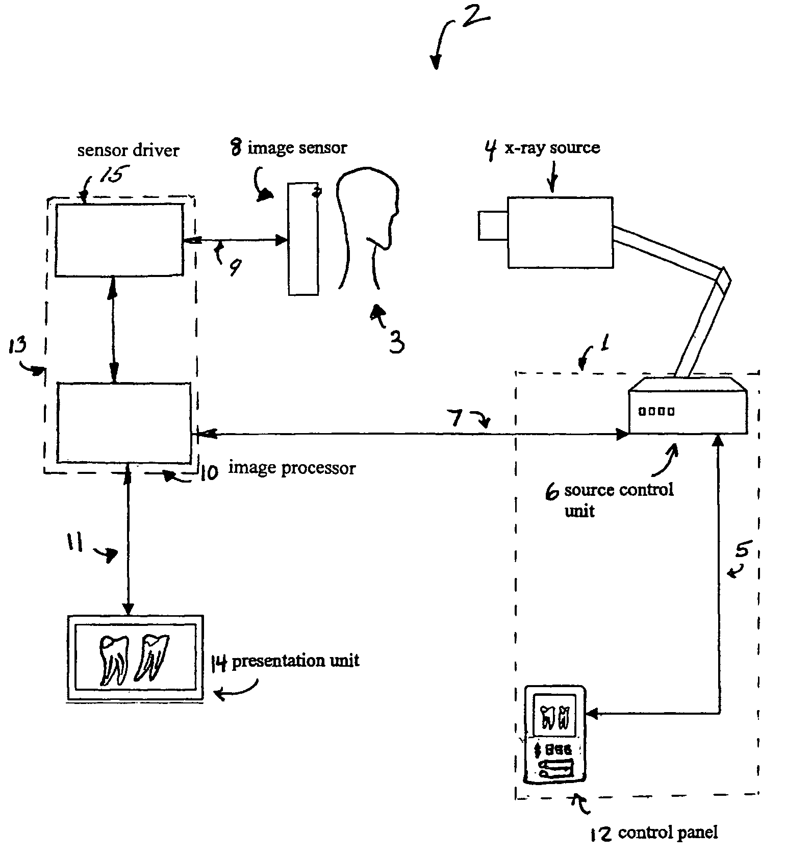
[0023]The present invention discloses a system and method for efficiently integrating x-ray generation and detection techniques. The system provides the ability to calculate and communicate the optimal radiation settings of a certain category and opacity of tooth in consideration of the specific physical anatomy of the subject patient. The system provides an arrangement whereby each component is electronically coupled to its corresponding functional mate providing for efficient signal transduction and communication of the exposure times to both the image sensor and x-ray source thereby coordinating their respective activities without the need for separate radiation sensing components. The system further provides the ability to view and manipulate the captured images at the point of x-ray source initiation.

[0024]In the following description, for purposes of explanation, numerous details are set forth in order to provide a thorough understanding of the present invention. However, it w...


PUM


Abstract

An integrated digital x-ray diagnostic system and a method for conducting dental radiography which includes optimizing exposure settings based on certain physical parameters of a patient, communicating the operational status of the x-ray generator and the exposure settings directly to the a CCD sensor without radiation sensitive detector elements, and supplying various viewing stations with image data thereby improving dental office workflow. The activation of the sensor and the x-ray source are coordinated so as to avoid pre-integration of charge in the image sensor, and at the same time reduce risk of over-exposure to the patient.

Description

BACKGROUND OF THE INVENTION[0001]The present invention relates to an integrated digital x-ray diagnostic system and a method for conducting dental radiography which includes optimizing exposure settings based on certain physical parameters of a patient, communicating the operational status of the x-ray generator and the exposure settings directly to a CCD sensor without radiation sensitive detector elements, and supplying various viewing stations with image data thereby improving dental office workflow.[0002]Digital image detectors, such as CCD sensors used in conventional video cameras, have been adapted to be x-ray sensitive elements in dental x-ray applications. For the past few decades, CCD sensors have been established as a leading technology in high-performance digital x-ray imaging. Unlike matrix-addressed sensors like amorphous silicon panels and CMOS imagers, CCD sensors read out their signal by transporting charge packets across their silicon substrate. The advantages of u...

Claims


Application Information

Patent Timeline
IPC IPC(8): H05G1/64
CPCA61B6/145A61B6/465A61B6/544A61B6/467H04N5/32A61B6/468A61B6/512H04N23/30
Inventor KREMA, ALAN P.STANTCHEV, GEORGECEKOV, LYUBOMIRGREENWOOD, MARK
Owner MIDMARK
Learn More