Regeneration and augmentation of bone using mesenchymal stem cells
a technology of mesenchymal stem cells and bone augmentation, which is applied in the field of bone augmentation and bone augmentation using mesenchymal stem cells, can solve the problems of large segmental defect repair in diaphyseal bone, significant morbidity, pain, and loss of function, and lack of osteoinduction compromise the desirability of bon
- Summary
- Abstract
- Description
- Claims
- Application Information
AI Technical Summary
Benefits of technology
Problems solved by technology
Method used
Image
Examples
example 1
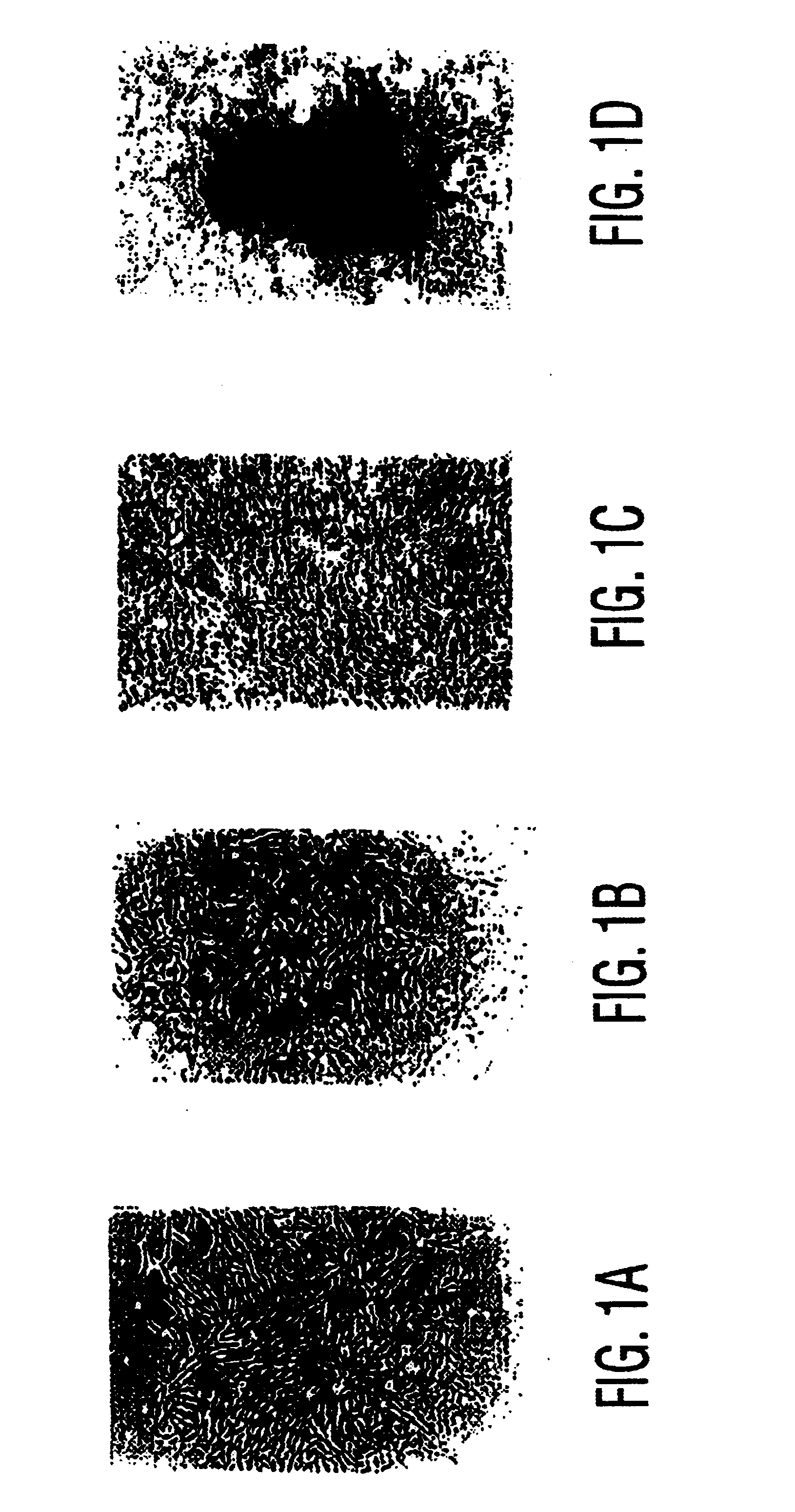
Rat Gap Defect Repair
Materials & Methods
Materials
[0064]Dexamethasone (Dex), sodium β-glycerophosphate (βGP), antibiotic penicillin / streptomycin, and alkaline phosphatase histochemistry kit #85 were purchased from Sigma Chemical Co. (St. Louis, Mo.), DMEM-LG (DMEM) tissue culture medium from GIBCO Laboratories (Grand Island, N.Y.), and L-ascorbic acid-2-phosphate (AsAP) from Wako Chemical (Osaka, Japan). Fetal bovine serum (FBS) was purchased from GIBCO following an extensive testing and selection protocol (35). Porous hydroxyapatite / β-tricalcium phosphate (HA / TCP) ceramic, mean pore size 200-450 βm, was generously provided by Zimmer, Inc. (Warsaw, Ind.). All other routine reagents used were of analytical grade.
MSC Isolation and Cultivation
[0065]MSC isolation and culture expansion was performed according to previously published methods. Briefly, male Fisher F344 rats (200-275 g) were sacrificed by pentobarbital overdose. The tibias and the femurs were recovered by dissection under st...
example 2
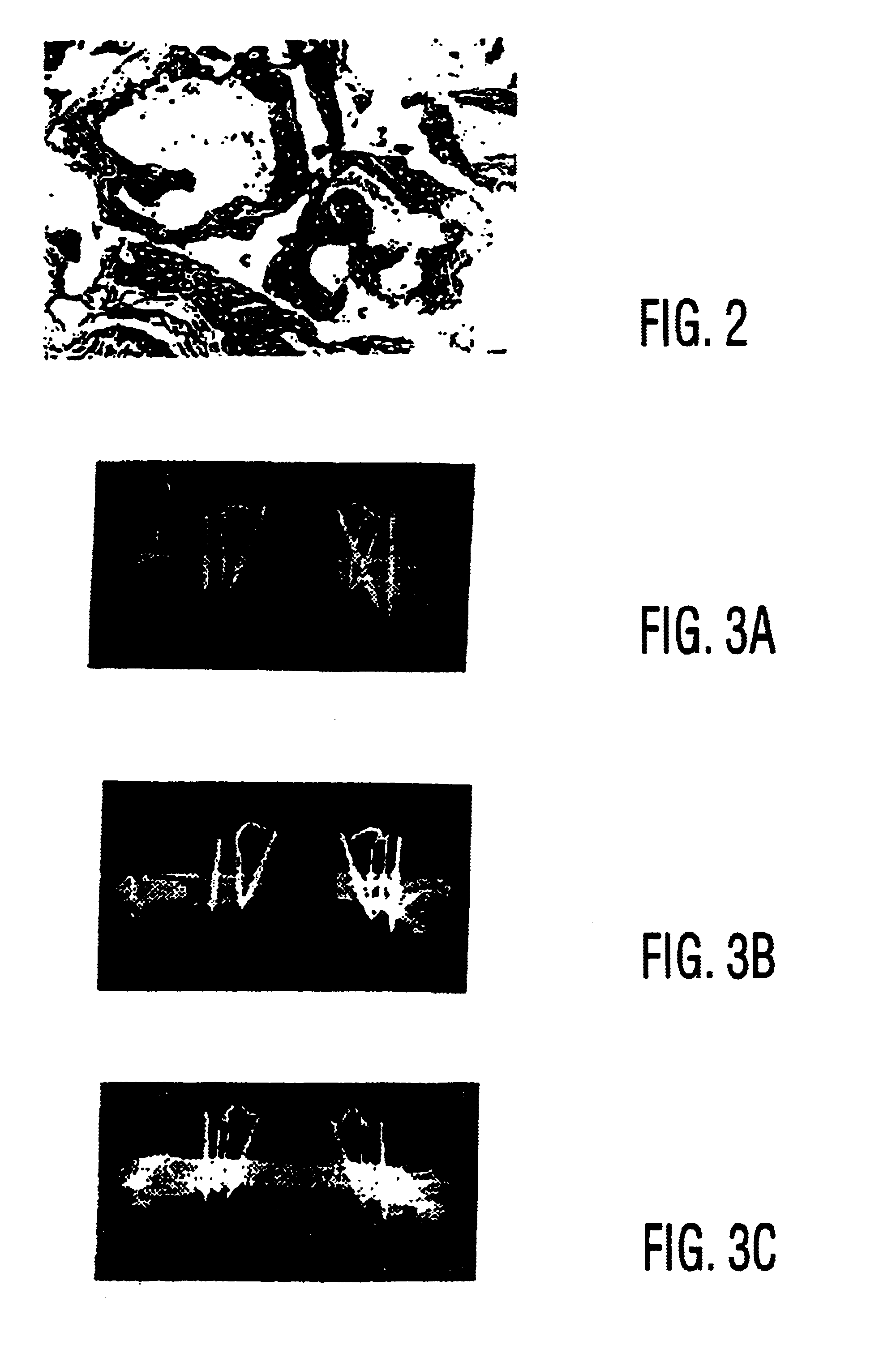
Large Segmental Canine Femoral Defects are Healed with Autologous Mesenchymal Stem Cell Therapy
[0090]This study demonstrates that culture-expanded, autologous mesenchymal stem cells can regenerate clinically significant bone defects in a large animal model.
[0091]Recently, the ability of syngeneic bone marrow-derived mesenchymal stem cells (MSCs) to repair large segmental defects in rodents was established (25). These MSCs may be isolated from marrow or periosteum, expanded in number ex vivo, and delivered back to the host in an appropriate carrier vehicle. Studies in rats demonstrated that the amount of bone formed 8 weeks following implantation of MSCs was twice that resulting from BMP delivered in the same carrier (25,50). In order to demonstrate clinical feasibility of this technology, our objective was to regenerate segmental bone defects in a large animal amenable to stringent biomechanical testing. To achieve this goal, we developed a canine femoral gap model to compare radiog...
example 3
in Vivo Bone Formation using Human Mesenchymal Stem Cells
[0100]Although rat MSCs have been shown to synthesize structurally competent bone in an orthotopic site (25), human MSCs have only been shown to form bone in vitro (2,23) and in an ectopic implantation site in immunodeficient mice. Since fracture healing and bone repair depend on the ability to amass enough cells at the defect site to form a repair blastema, one therapeutic strategy is to directly administer the precursor cells to the site in need of repair. This approach is particularly attractive for patients who have fractures which are difficult to heal, or patients who have a decline in their MSC repository as a result of age (28,47), osteoporosis (5 1), or other metabolic derangement. With this in mind, the goal of the current study was to show that purified, culture-expanded human MSCs are capable of regenerating bone at the site of a clinically significant defect.
Materials and Methods
Human MSC Cultivation and Manipulat...
PUM
| Property | Measurement | Unit |
|---|---|---|
| Time | aaaaa | aaaaa |
| Time | aaaaa | aaaaa |
| Composition | aaaaa | aaaaa |
Abstract
Description
Claims
Application Information
 - R&D
- Intellectual Property
- Life Sciences
- Materials
- Tech Scout
- Unparalleled Data Quality
- Higher Quality Content
- 60% Fewer Hallucinations
Browse by: Latest US Patents, China's latest patents, Technical Efficacy Thesaurus, Application Domain, Technology Topic, Popular Technical Reports.
© 2025 PatSnap. All rights reserved.Legal|Privacy policy|Modern Slavery Act Transparency Statement|Sitemap|About US| Contact US: help@patsnap.com


