
Α-conotoxin peptides

a technology of conotoxin and peptide, which is applied in the direction of peptides, peptide/protein ingredients, peptide sources, etc., can solve the problems of long duration of action of nondepolarizing agents, no siginificant affinity of neuronal nicotinic receptors, etc., and achieves rapid recovery of drug effects, shortening the onset time without dramatic extension of recovery times

Inactive Publication Date: 2005-02-15
UNIV OF UTAH RES FOUND +1
View PDF14 Cites 16 Cited by
  • Summary
  • Abstract
  • Description
  • Claims
  • Application Information

AI Technical Summary

Benefits of technology

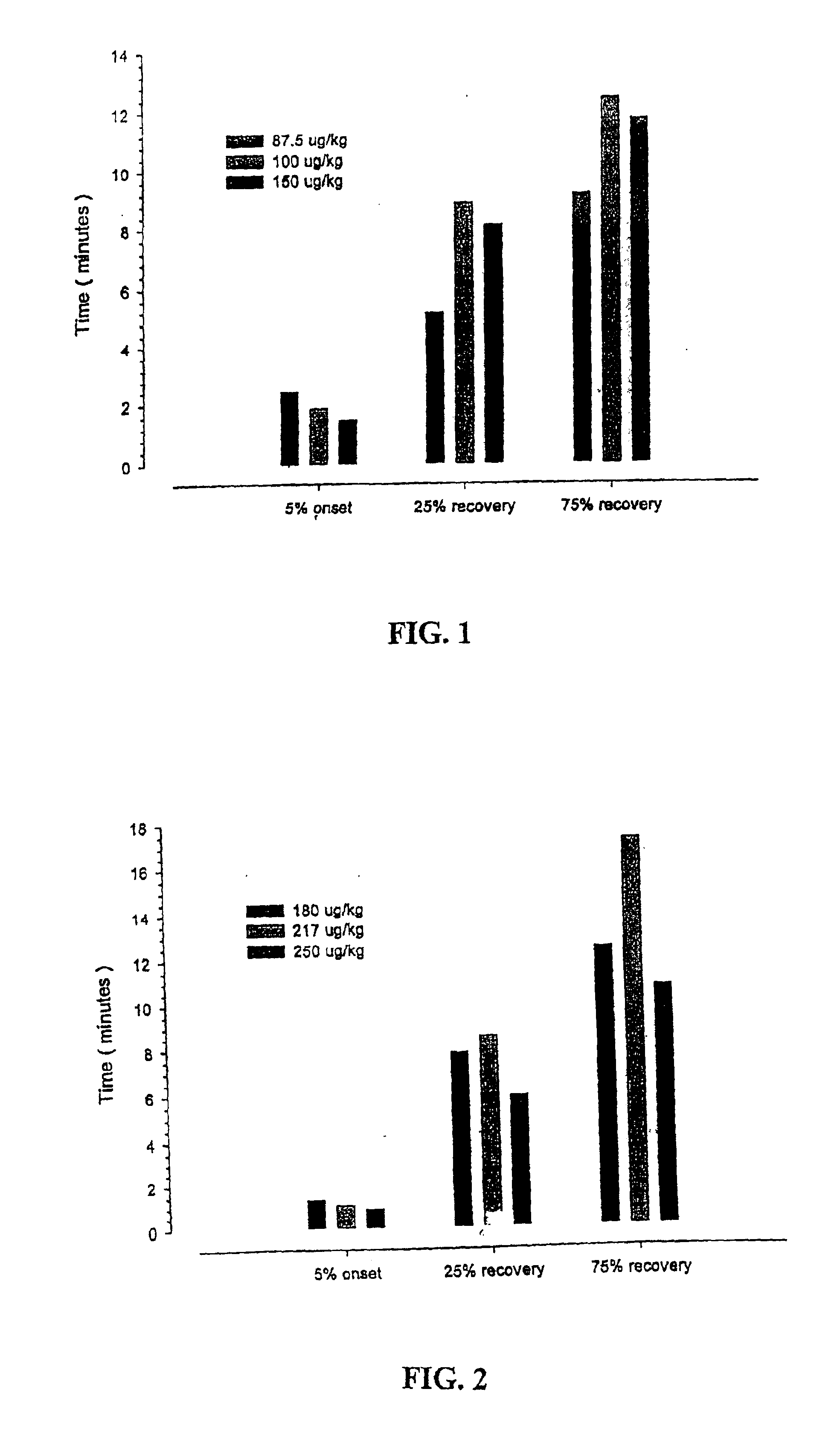
[0153]This study was an open label, dose-ranging, single center investigation. A total of 14 rats were studied (10 in each of five groups). All animals were anesthetized with pentobarbital (60 mg/kg) given by intraperitoneal administration and maintained with supplemental doses as determined by physiological monitoring variables. A tracheotomy was performed and the rats were ventilated with room air keeping PCO2 near 35 torr. The carotid artery was cannulated to measure blood pressure and arterial blood gases. The right jugular vein was cannulated for intravenous infusion and further drug administration. Body temperature was maintained at 36°-38° C. during the entire experiment. The sciatic nerve was exposed in the popliteal space and stimulated with train-of-four stimulation using a Digistim nerve stimulator. The tivialis anterior muscle contractoin was measured by attaching the rat hind limb to an isometric force transducer to record the evoked response. Prior to administration of the study drug, baseline measurements of blood pressure, heart rate and muscle contraction force were measured for a five-minute period and at five minute intervals for the duration of the study.
[0154]The initial dose for analysis was based on biologically effective doses determined in mice. Based on the onset, maximum effect and duration of effect from the first animal studied, the dose for the next animal was either doubled or halved. If the relaxation level was maintained at a maximal level for greater than 20 minutes from this initial dose, then the subsequent dose studied was doubled. this progression continued until the dose that produced near maximal muscle relaxation was found.
[0155]The conopeptide derivatives MI and GI were studied in the initial study. For each compound

Problems solved by technology

However, none of these peptides show siginificant affinity for neuronal nicotinic receptors.
The long duration of action of nondepolarizing agents is unacceptable in many surgical procedures which take less than one hour because the patient is not generally filly recovered from their effects e.g., the patient may be unable to breathe adequately on his or her own.
While such drugs can be pharmacologically antagonized with anticholinesterase agents, this obviously necessitates the administration of a second drug which itself may have its own side effects e.g., bradycardia, gut spasm and bronchorrhea.

Method used


Image

  • Α-conotoxin peptides
  • Α-conotoxin peptides
  • Α-conotoxin peptides

Examples


example 1

Dose-Effect Study for MI and GI

[0153]This study was an open label, dose-ranging, single center investigation. A total of 14 rats were studied (10 in each of five groups). All animals were anesthetized with pentobarbital (60 mg / kg) given by intraperitoneal administration and maintained with supplemental doses as determined by physiological monitoring variables. A tracheotomy was performed and the rats were ventilated with room air keeping PCO2 near 35 torr. The carotid artery was cannulated to measure blood pressure and arterial blood gases. The right jugular vein was cannulated for intravenous infusion and further drug administration. Body temperature was maintained at 36°-38° C. during the entire experiment. The sciatic nerve was exposed in the popliteal space and stimulated with train-of-four stimulation using a Digistim nerve stimulator. The tivialis anterior muscle contractoin was measured by attaching the rat hind limb to an isometric force transducer to record the evoked respo...

example 2

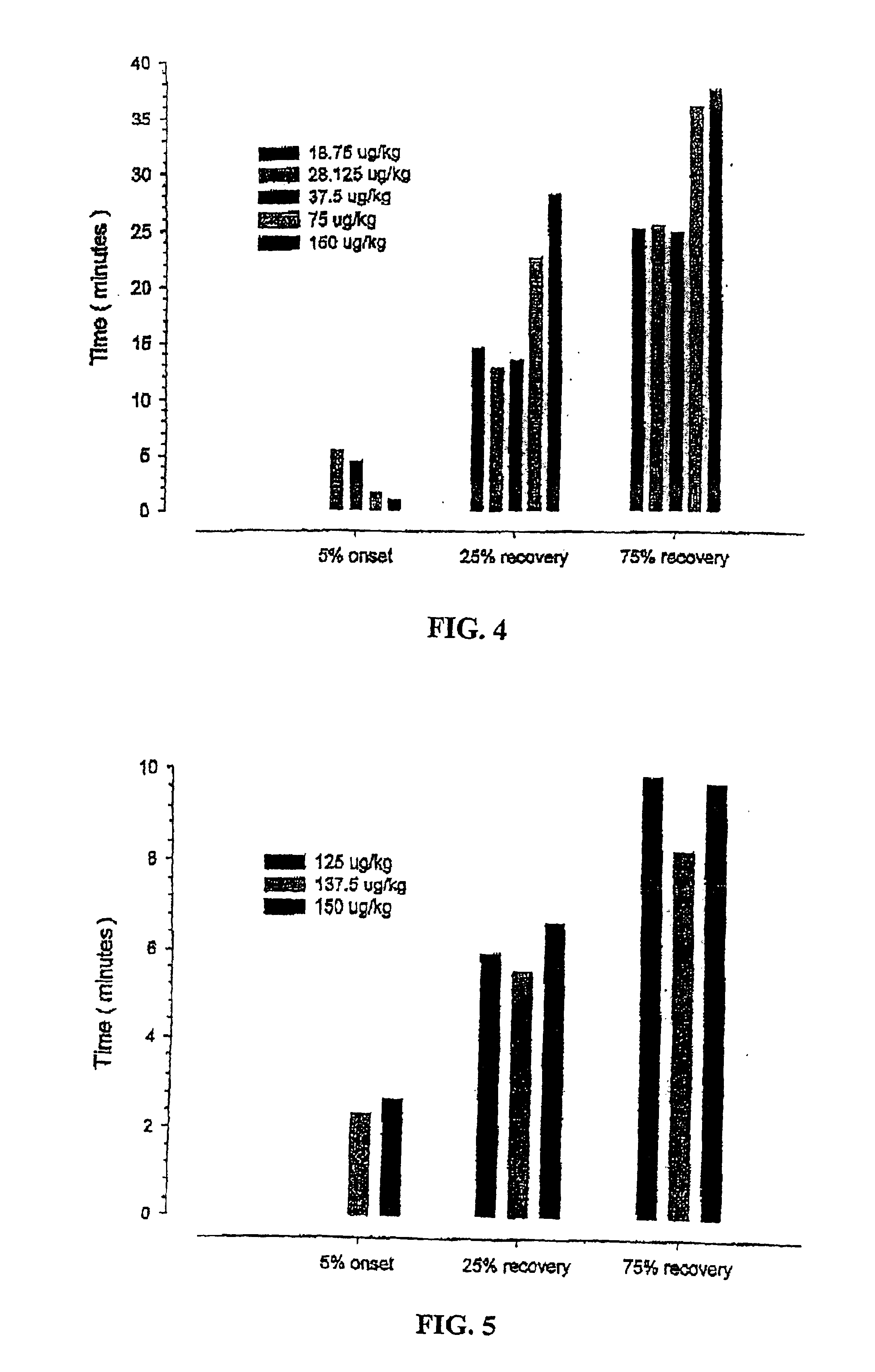
Dose-Effect Study for Iodinated-MI

[0161]A similar study as described in Example 1 was conducted for two iodinated derivatives of MI, namely, mono-iodo-Tyr12-MI and di-iodo-Tyr12-MI. The onset and recovery results for mono-iodo-Tyr12-MI and di-iodo-Tyr12-MI are shown in FIGS. 4 and 5, respectively. Dose-response plots for mono-iodo-Tyr12-MI and di-iodo-Tyr12-MI were made to estimate the ED50 dose of these agents. The ED50 values are ˜16 μg / kg for mono-iodo-Tyr12-MI and ˜92.5 μg / kg for di-iodo-Tyr12-MI.

example 3

Muscle Relaxant Effect in Anesthetized Monkeys

[0162]The peptides MI, GI, EI, mono-iodo-MI and di-iodo-MI are each separately dissolved 0.9 percent saline at a concentration of 2 mg / ml. Rhesus monkeys are anesthetized with halothane, nitrous oxide and oxygen. The maintenance concentration of halothane is 1.0%. Arterial and venous catheters are placed in the femoral vessels for drug administration and recording of the arterial pressure. Controlled ventilation is accomplished via an endotrachael tube. Twitch and tetanic contractions of the tibialis arterior muscle are elicited indirectly via the sciatic nerve. Recordings of arterial pressure electrocardiogram (lead I), heart rate, and muscle function are made simultaneously. Four to six animals received each listed compound. Four additional animals received succinylcholine chloride or d-tubocurarine chloride as controls. Is is seen that the tested compounds generally provide similar or better results than those seen for succinylcholine...


PUM

PropertyMeasurementUnit
Timeaaaaaaaaaa
Timeaaaaaaaaaa
Timeaaaaaaaaaa

Abstract

The invention relates to relatively short peptides (termed α-conotoxins herein), about 10-25 residues in length, which are naturally available in minute amounts in the venom of the cone snails or analogous to the naturally available peptides, and which preferably include two disulfide bonds. The α-conotoxins, as described herein, are useful for as neuromuscular blocking agents, such as muscle relaxants.

Description

CROSS-REFERENCE TO RELATED APPLICATIONS[0002]The present application is a continuation-in-part of patent application Ser. No. 09 / 488,799 filed on 21 Jan. 2001 now U.S. Pat. No. 6,268,473 and claims benefit thereto. The present application also claims benefit under 35 USC §119(e) to U.S. provisional patent applications Ser. No. 60 / 116,881 filed on 22 Jan. 1999, Ser. No. U.S. 60 / 116,882 filed on 22 Jan. 1999, 60 / 219,407 filed on 20 Jul. 2000 and Ser. No. 60 / 221,557 filed on 28 Jul. 2000. Each of these applications is incorporated herein by reference.[0003]This invention was made with Government support under Grant No. PO1 GM48677 awarded by the National Institute of General Medical Sciences, National Institutes of Health, Bethesda, Md. and under SBIR grant No. 1 R43 GM62064-01. The United States Government has certain rights in the invention.BACKGROUND OF THE INVENTION[0004]The invention relates to relatively short peptides (termed α-conotoxins herein), about 10-25 residues in length,...

Claims


Application Information

Patent Timeline
IPC IPC(8): A61K38/17C07K14/435A61K38/00
CPCC07K14/43504A61K38/00
Inventor OLIVERA, BALDOMERO M.LAYER, RICHARD T.MCINTOSH, J. MICHAELNIELSEN, JACOB SCOTTJONES, ROBERT M.
Owner UNIV OF UTAH RES FOUND
Learn More