Antibody combinations for treatment of cancer in specific patients
a cancer and specific patient technology, applied in the field of specific patient antibody combinations, can solve the problems of unresponsiveness and resistance to pd-1/pd-l1 antibodies, no longer benefiting from treatment, and presenting a significant burden on payers and health care systems
- Summary
- Abstract
- Description
- Claims
- Application Information
AI Technical Summary
Benefits of technology
Problems solved by technology
Method used
Image

Examples
example 1
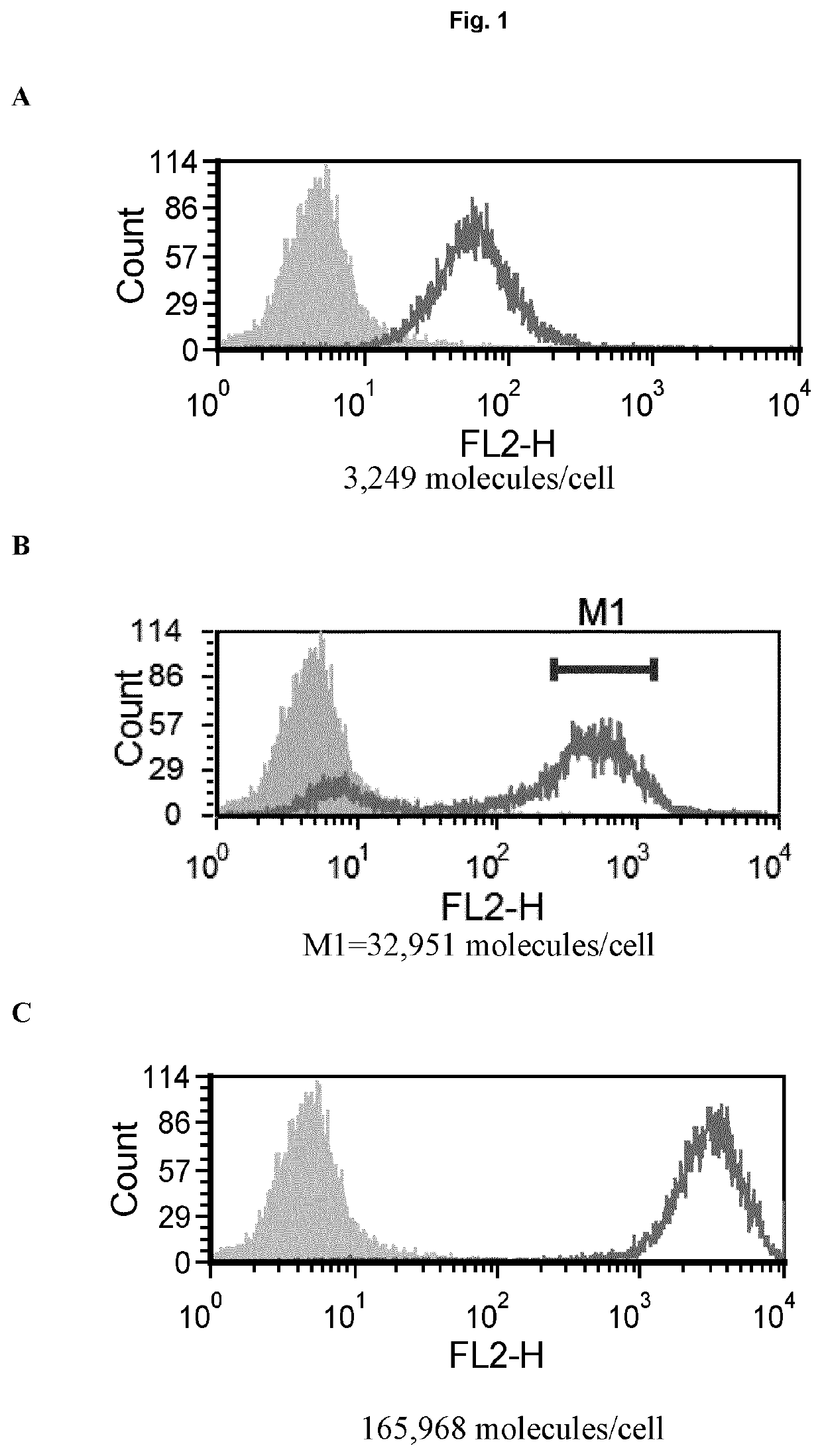
[0154]For transfection of Jurkat cells, the cells were cultured in RPMI-1640 medium containing 10% fetal calf serum, FCS (Sigma), L-glutamine (Life Technologies), sodium pyruvate (Life Technologies) and Pen-Strep (Life Technologies). The day before transfection, the cells were split to 0.5×10⁶ / ml and cultured over night. To transfect the cells, 1×10⁶ cells were centrifuged at 90×G for 10 minutes and thereafter resuspended in 100 μl nucleofector solution (Amaxa® Cell Line Nucleofector® Kit V, Lonza) where 2 μg DNA (hPD-1 in pcDNA3) was added. The mixture was then transferred to a nucleofector cuvette. Cuvettes were placed in nucleofector II machine and nucleofected with program X-005. After incubation at room temperature (around 18-22° C.) for 10 min, 500 ml media was added to the cuvette and transferred to a 12-well plate containing 1 ml media. To select for transfected cells, geneticin was added at 1 mg / ml 48 hours after transfection. 10-14 days later, p...
example 2
tification on Immune Cells from Tumor-Bearing Mice
[0164]PD-1 receptor numbers on immune cells from mice tumors were determined using Quantum™ Simply Cellular® beads (Bangs Laboratories, Inc.). In brief, beads were stained with rat anti PD-1 antibody (clone 29F.1A12, BioLegend) to create a standard curve. Cell samples were then read against the curve for determination of expression.
[0165]Quantification was done on cells from tumor-bearing mice. Mice were bred and maintained in local facilities in accordance with home office guidelines. Six to eight weeks-old female C57 / BL6 mice were supplied by Taconic (Bomholt, Denmark) and maintained in local animal facilities. MC38 cells (ATCC) were grown in glutamax buffered RPMI supplemented with 10% FBS. When cells were semi confluent they were detached with trypsin and re-suspended in sterile PBS at 10×106 cells / ml. Mice were s.c. injected with 100 μl cell suspension corresponding to 1×10⁶ cells / mouse. Tumors were grown for ˜20 days before col...
example 3
onal Effect with Anti PD-1 In-Vivo
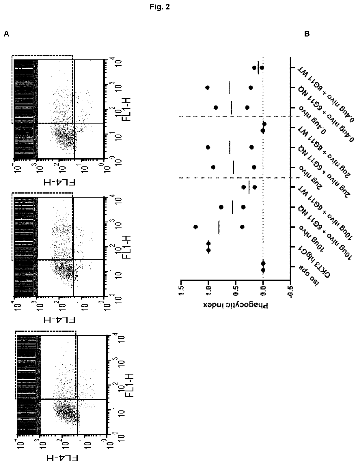
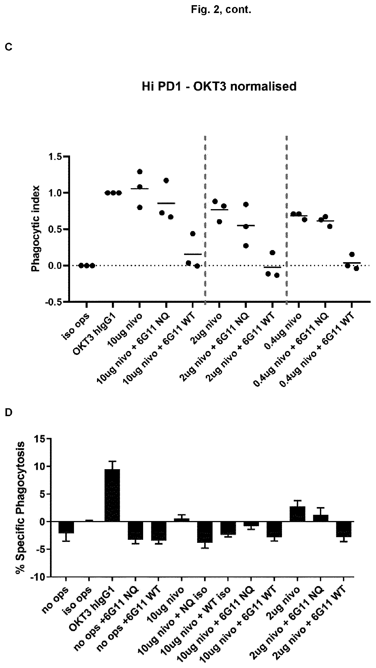
[0166]The PD-1 expression on mice cells correspond to expression levels between ‘mid and high’ on transfected Jurkat cells (Example 1, FIG. 1). In a phagocytosis assay, BI-1206 was shown to significantly reduce the level of phagocytosis for these ‘mid and high’ PD-1 expressing cells (Example 1, FIG. 2). This data, in combination with the improved therapeutic anti-tumor effect seen when combining anti PD-1 with anti-FcγRIIb (BI-1206 mouse surrogate) in the MC38 model in-vivo (FIG. 3), suggests an improved therapeutic effect of anti PD-1 in combination with BI-1206 in patients with a medium or high PD-1 expression, i.e. a PD-1 expression that is equal to or higher than 15,500 PD-1 molecules / cell.
PUM
| Property | Measurement | Unit |
|---|---|---|
| concentration | aaaaa | aaaaa |
| concentration | aaaaa | aaaaa |
| concentration | aaaaa | aaaaa |
Abstract
Description
Claims
Application Information
 - R&D
- Intellectual Property
- Life Sciences
- Materials
- Tech Scout
- Unparalleled Data Quality
- Higher Quality Content
- 60% Fewer Hallucinations
Browse by: Latest US Patents, China's latest patents, Technical Efficacy Thesaurus, Application Domain, Technology Topic, Popular Technical Reports.
© 2025 PatSnap. All rights reserved.Legal|Privacy policy|Modern Slavery Act Transparency Statement|Sitemap|About US| Contact US: help@patsnap.com