
Engineered Intestinal Tissue and Uses Thereof

a technology of intestinal tissue and engineering, applied in the field of intestinal tissue models, can solve the problems of low safety and efficacy predictability, drug development attrition, current preclinical models are limited in their ability to capture, and systemic availability

Pending Publication Date: 2022-06-30
ORGANOVO
View PDF0 Cites 0 Cited by
  • Summary
  • Abstract
  • Description
  • Claims
  • Application Information

AI Technical Summary

Benefits of technology

[0011]The present invention provides intestinal tissue models that offer advantages over existing in vitro assay systems by providing an intestinal epithelial cell layer on top of a layer of intestinal interstitial tissue comprising myofibroblasts and, optionally, other key cell types such as myeloid immune cells, smooth muscle cells, endothelial cells and neurons. These intestinal tissue models allow one to see the impact of treatments in a more holistic system. And, the intestinal tissue models support epithelial morphology and function for an extended period of time in culture, thus enabling chronic studies of treatments. Because of their multi-cellularity and architecture, the intestinal tissue constructs provide a unique system to study multi-faceted processes including secretion, transport, cell-cell interactions and pathogenic processes, including inflammation and cancer.
[0013]The intestinal tissue models described herein provide an opportunity to accurately study how compounds affect the intestinal tissue as well as to model pathogenic processes in the intestine. The intestinal tissue models disclosed herein are useful for predicting toxicity of pharmaceutical compounds earlier in the drug development process. By incorporating both diseased (ex: inflamed or tumor tissue) and normal cell compartments into the same tissue, the impact of therapeutic agents on both healthy and diseased tissue can be assessed in the same tissue system. Intestinal tissue constructs comprising primary cells are especially useful for personalized medicine.
[0044]In some embodiments, the phenotype of an intestinal disorder or injury is induced by contacting the intestinal tissue model with a treatment, compound, or infectious agent that gives rise to the phenotype. In some embodiments, the phenotype of an intestinal disorder or injury is the presence of tumor(s), tumor fragment(s), tumor cells, or immortalized cells in the intestinal tissue model. In some embodiments, the ability of a candidate therapeutic agent to reverse, reduce, induce or prevent an intestinal disorder or injury is reduced tumor(s), tumor fragment(s), tumor cells, or immortalized cells invasion or metastasis.

Problems solved by technology

The intestinal mucosa plays a crucial role in regulating absorption, first-pass metabolism, clearance, drug-drug interactions, and can be a site of drug induced toxicity.
Current in vitro systems and preclinical models utilized in drug development do not adequately recapitulate the complexities of native human intestinal tissue, leading to low safety and efficacy predictability and attrition in drug development.
Current preclinical models are limited in their ability to capture the complexities and function of human intestinal tissue [1-5].
Systemic availability, diminished efficacy, and off target effects remain challenges to the successful prediction of candidate drugs and contribute to attrition in drug development.
Many predictive challenges can be attributed to the lack of preclinical tools to model the complexities of intestinal function in vitro [1-3].
Standard 2D systems lack the complexity to accurately model outcomes such as low bioavailability, the result of a combination of low permeability and interplay of metabolic enzymes with influx and efflux transporters in the intestinal epithelium [3].
Microsomes are a convenient tool for the initial assessment of metabolism, but cannot model cellular level outcomes.
2D cell monolayer models lack native context of cell-cell and cell-matrix interactions and are phenotypically limited, while genetic disparity of animal models may not provide a high correlation with human outcomes [16].
However, altered metabolism in tumor cells compared to normal tissue is a major disadvantage of these models.
And, these tumor models do not represent features of native intestinal epithelium.
Limitations of the Caco-2 model include a lack of P-450 metabolizing enzyme expression and activity, lack of robust intestinal transporter expression and function, variation with passage number, and inconsistencies between clones in the line.
Monocultures of primary intestinal epithelial cells more closely resemble in vivo tissue but may have limited intestinal epithelial function in part to their isolation from the other supportive cell types present in the intestinal wall.
These structures suffer from low availability (from humans), limited viable lifespan in vitro, and may lack in vivo organ physiology.
Notably, the closed lumens and the inward orientation of epithelia in intestinal organoids makes the apical surface relatively inaccessible for the direct stimulation they would normally experience in vivo, and makes organoids incompatible with most standard ADMF / Tox assays.
Intestinal slices, however, have limited viability ex vivo and only function for about 24 hours.
Furthermore, these tissues are not compatible with cryopreservation, which limits their use to short term studies [15].
Animal models are frequently used to estimate compound bioavailability, however genetic differences can lead to disparity in expression of metabolic enzymes and transporters compared to humans which can result in poor prediction [2, 3, 16].

Method used


Image

  • Engineered Intestinal Tissue and Uses Thereof
  • Engineered Intestinal Tissue and Uses Thereof
  • Engineered Intestinal Tissue and Uses Thereof

Examples


example 1

A Manually Created Three-Dimensional Intestinal Tissue Model

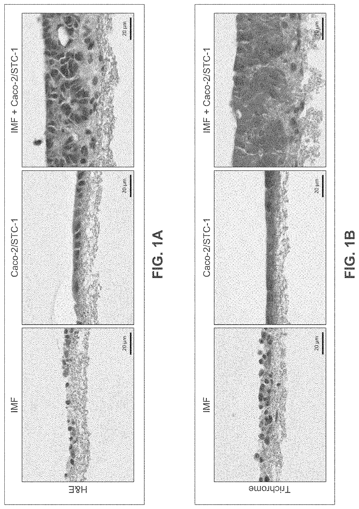
[0288]Human intestinal tissue was fabricated with human primary intestinal cells and intestinal cell lines by continuous deposition using interstitial bio-ink containing gelatin and manual deposition of an epithelial suspension.

[0289]Bio-ink was generated by a cellular mixture of 100% primary adult human intestinal myofibroblasts (IMF) in 8% gelatin in a concentration of 20 million cells per milliliter. Three-dimensional bioink constructs were printed by continuous deposition using the Novogen MMX Bioprinter®. One tissue was printed per transwell in a 24 well plate. The transwell printing surface contained a polytetrafluoroethylene (PTFE) membrane coated with equimolar mixture of types I and III collagen (bovine) with pores 3 μm in size. Following printing, tissues were allowed to mature for 4 days in a humidified incubator in growth media. Tissues were cultured in 100% IMF media and media was changed daily. After incubatio...

example 2

A Bioprinted Three-Dimensional Intestinal Tissue Model

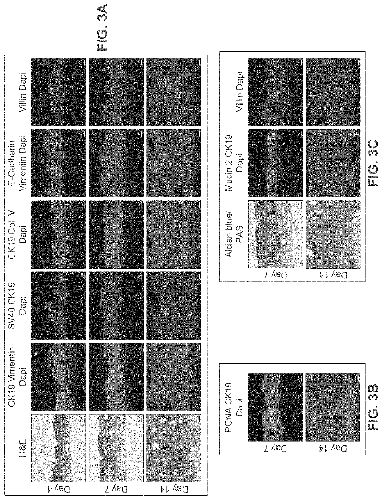
[0297]A human intestinal tissue construct was fabricated by bioprinting with 100% human adult primary intestinal cells by continuous deposition using interstitial bio-ink containing collagen followed by deposition of epithelial suspension.

[0298]Bio-ink was generated by a cellular mixture of 100% primary adult human intestinal myofibroblasts (IMF) in 100% bovine type I collagen at a concentration of 20 million cells per milliliter. Three-dimensional bio-ink constructs were printed by continuous deposition using the Novogen MMX Bioprinter® in a base layer to create an interstitial structure. One tissue was printed per transwell in a 24 well plate. The transwell printing surface contained a polytetrafluoroethylene (PTFE) membrane coated with equimolar mixture of types I and III collagen (bovine) with pores 3 um in size. Following printing, tissues were allowed to mature for 4 days in a humidified 37° C. incubator in 100% IMF media a...

example 3

A Bioprinted Three-Dimensional Intestinal Tissue Model Comprising Enteroendocrine Cells

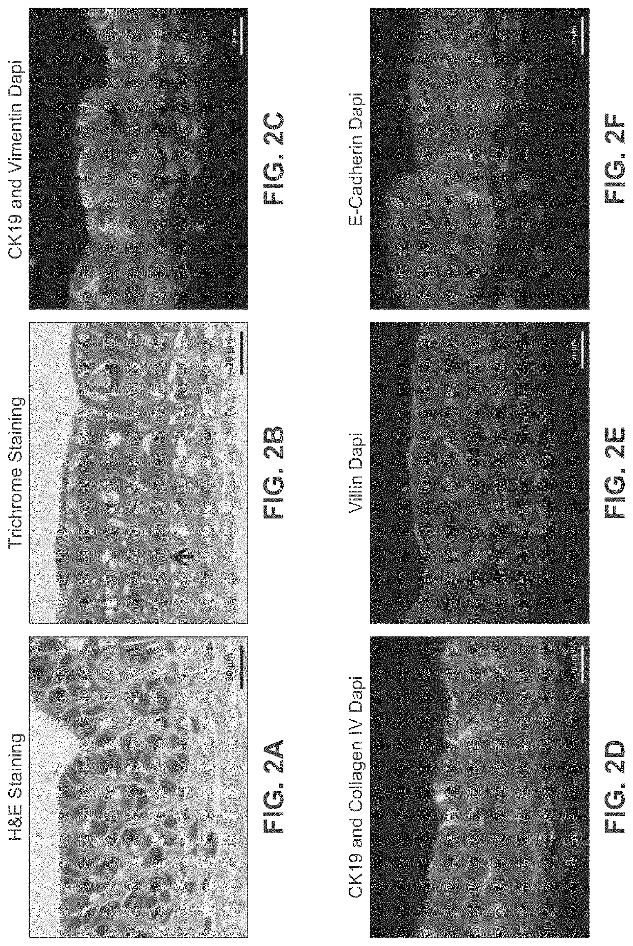
[0307]A human intestinal tissue construct was fabricated with human adult primary intestinal cells and mouse enteroendocrine cell line STC-1 by continuous deposition using an interstitial bio-ink containing collagen and manual deposition of an epithelial suspension.

[0308]The interstitial layer was generated in an identical manner to Example 2. Bio-ink was generated by a cellular mixture of 100% primary adult human intestinal myofibroblasts (IMF) in bovine type I collagen at a concentration of 20 million cells per milliliter. Three-dimensional bioink constructs were printed by continuous deposition using the Novogen MMX Bioprinter® in a base layer with to create an interstitial structure. One tissue was printed per transwell in a 24 well plate. The transwell printing surface contained a polytetrafluoroethylene (PTFE) membrane coated with equimolar mixture of types 1 and III collagen (bovine) with p...


PUM

PropertyMeasurementUnit
timeaaaaaaaaaa
thickaaaaaaaaaa
thickaaaaaaaaaa

Abstract

Disclosed are methods of assessing the ability of a candidate therapeutic agent to reverse, reduce or prevent intestinal injury by a potential toxic agent using a three-dimensional, engineered, bioprinted, biological intestinal tissue model. Also disclosed are methods of assessing the effect of an agent on intestinal function, the method comprising contacting the agent with a three-dimensional, engineered, bioprinted, biological intestinal tissue model.

Description

BACKGROUND OF THE INVENTIONField of the Invention[0001]The invention is in the field of intestinal tissue models and their use in assays. Disclosed are methods of assessing the ability of a candidate therapeutic agent to reverse, reduce or prevent intestinal injury by a potential toxic agent using a three-dimensional, engineered, bioprinted, biological intestinal tissue model. Also disclosed are methods of assessing the effect of an agent on intestinal function, the method comprising contacting the agent with a three-dimensional, engineered, bioprinted, biological intestinal tissue model.Background Art[0002]The intestinal mucosa plays a crucial role in regulating absorption, first-pass metabolism, clearance, drug-drug interactions, and can be a site of drug induced toxicity. Current in vitro systems and preclinical models utilized in drug development do not adequately recapitulate the complexities of native human intestinal tissue, leading to low safety and efficacy predictability a...

Claims


Application Information

Patent Timeline
Patent Type & Authority Applications(United States)
IPC IPC(8): C12N5/071A61K35/15G01N33/50A61K35/38
CPCC12N5/0679A61K35/15G01N33/5088A61K35/38C12N2535/00C12N2502/23C12N2513/00C12N2533/30C12N2533/54C12N2502/1335C12N5/0697A61P1/00A61P35/00A61P3/06A61P3/10A61P9/12A61K35/12A01K67/027A01K2207/12A01K2267/03C12N5/0634
Inventor RETTING, KELSEY NICOLENGUYEN, DEBORAH LYNN GREENEMADDEN, LAURAN
Owner ORGANOVO
Learn More