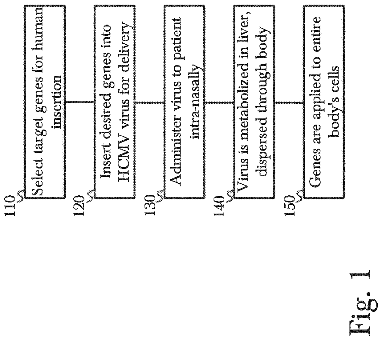
Gene therapy using genetically modified viral vectors
a technology of genetically modified viruses and gene therapy, applied in the field of gene therapy, can solve the problems of raising the already lofty cost of gene therapy, unable to commercially viable method to extend the telomeres at the end of chromosomes in human cells, and exceedingly expensive gene therapy for patients, etc., to achieve effective, long-lasting, and convenient insertion into patients.
- Summary
- Abstract
- Description
- Claims
- Application Information
AI Technical Summary
Benefits of technology
Problems solved by technology
Method used
Image

Examples
example 1
dia, and Viruses
[0163]An MCMV bacterial artificial chromosome (MCMV-BAC) Smith strain was used. Mouse fibroblast 3T3 cells were used for the MCMV culture and growth assays and were cultured in minimal essential media (MEM) with 10% fetal bovine serum (FBS), 1% penicillin and streptomycin (P / S) at 37° C., 5% CO2. Approved Institutional Biosafety Committee (IBC) and IACUC protocol were followed.
Example 2: Construction and Characterization of Recombinant MCMVLuc-TERT and MCMVLuc-FS344
[0164]Recently, the CMV vector has emerged as a potential delivery vector for expressing therapeutic molecules, including proteins. CMV can infect different cell types in the body and is thus able to deliver the target proteins to numerous cell types. More specifically, CMV can infect fibroblast, hepatocytes, endothelial cells, macrophages, epithelial cells, lymphocytes, retinal pigment epithelial cells, and cells of the gastrointestinal tract. Therefore, CMV can infect and deliver its target antigens to ...
example 4
ntolerance Test
[0170]Three mice from each group were selected at 22 months of age: (1) MCMVLuc-WT-IN, (2) MCMVLuc-WT-IP, (3) Uninfected, (4) MCMVLuc-TERT-IP, (5) MCMVLuc-TERT-IN, (6) MCMVLuc-FS344-IP, and (7) MCMVLuc-FS344-IN. The mice were starved for 15 hours, followed by intraperitoneal injection with a 50 mg glucose solution. Blood samples were collected at 0 min, 15 min, 30 min, 60 min, 120 min, 180 min, 240 min, 300 min, 360 min, 420 min, and 480 min via a small incision in the tail vein of the mice. The blood glucose level was immediately determined using OneTouch Ultra glucose strips.
[0171]A peak of glucose level was observed at 30 minutes of glucose administration (FIG. 22A). After 30 minutes, the uninfected or WT treated mice were showing high blood glucose with an average value of 310 mg / dl and 316 mg / dl respectively, whereas the mTERT and mFS344 treated mice were showing 154 mg / dl and 165 mg / dl glucose. Moreover, blood glucose level came back to the normal basal level in...
example 7
NA Isolation, cDNA Preparation and Real-Time PCR
[0177]To determine the level of mTERT and mFS344 in various tissues of mice infected with MCMVLuc-TERT, real-time PCR was performed on cDNA prepared from RNA of infected tissues. Eleven-month-old mice (3 in each group) were infected with wild type MCMV, MCMVLuc-TERT, and MCMVLuc-Fs344. The heart, brain, lung, liver, and kidney were isolated 6-days post infection. Uninfected mice were used as a control. The tissues were homogenized, and RNA was isolated using RNeasy® Mini Kit (Qiagen). Purified RNA concentration was determined by measuring optical density. Approximately, 1.0 μg of RNA was used to prepare cDNA using a Titanium RT-PCR kit (TaKaRa). Real-time PCR was performed on the cDNA using mTERT, and mFS344 primers as described previously. β-actin was used as an internal control for normalization. The expression of mTERT and mFS344 was determined in respective tissues after normalization with β-actin. Results for TERT mRNA levels are ...
PUM
| Property | Measurement | Unit |
|---|---|---|
| temperatures | aaaaa | aaaaa |
| time | aaaaa | aaaaa |
| time | aaaaa | aaaaa |
Abstract
Description
Claims
Application Information
 - R&D
- Intellectual Property
- Life Sciences
- Materials
- Tech Scout
- Unparalleled Data Quality
- Higher Quality Content
- 60% Fewer Hallucinations
Browse by: Latest US Patents, China's latest patents, Technical Efficacy Thesaurus, Application Domain, Technology Topic, Popular Technical Reports.
© 2025 PatSnap. All rights reserved.Legal|Privacy policy|Modern Slavery Act Transparency Statement|Sitemap|About US| Contact US: help@patsnap.com