
Small tunable fluorophores for the detection and imaging of biomolecules

a bioactive molecule and fluorophores technology, applied in the field of small, conjugatable, orthogonal and tunable fluorophores for imaging of small bioactive molecules, can solve the problems of inability to image in situ small metabolites in live cells and intact organisms, and inability to detect small bioactive molecules in the presence of living cells

Pending Publication Date: 2022-05-19
THE UNIV COURT OF THE UNIV OF EDINBURGH
View PDF0 Cites 0 Cited by
  • Summary
  • Abstract
  • Description
  • Claims
  • Application Information

AI Technical Summary

Benefits of technology

The present invention provides a collection of small, fluorescent molecules that can be used for various applications such as cancer cell labeling, immune cell labeling, stem cell labeling, and optical imaging modalities. These molecules have the advantage of being tunable, meaning they can be adjusted to emit light across the entire visible spectrum. Additionally, they can be used for other optical imaging modalities such as surface-enhanced Raman scattering and optical coherence tomography. The molecules have the ability to be detected under multiple imaging techniques and can be used for therapeutic, diagnostic, surgery, and analytical methods.

Problems solved by technology

Despite advances in fluorescence imaging to label small biomolecules, there are few approaches to image in situ small metabolites in live cells and intact organisms.
However, fluorescent labels, in particular red and near-infrared (NIR) fluorophores, are bulky structures that can impair how small biomolecules traffic within cells.
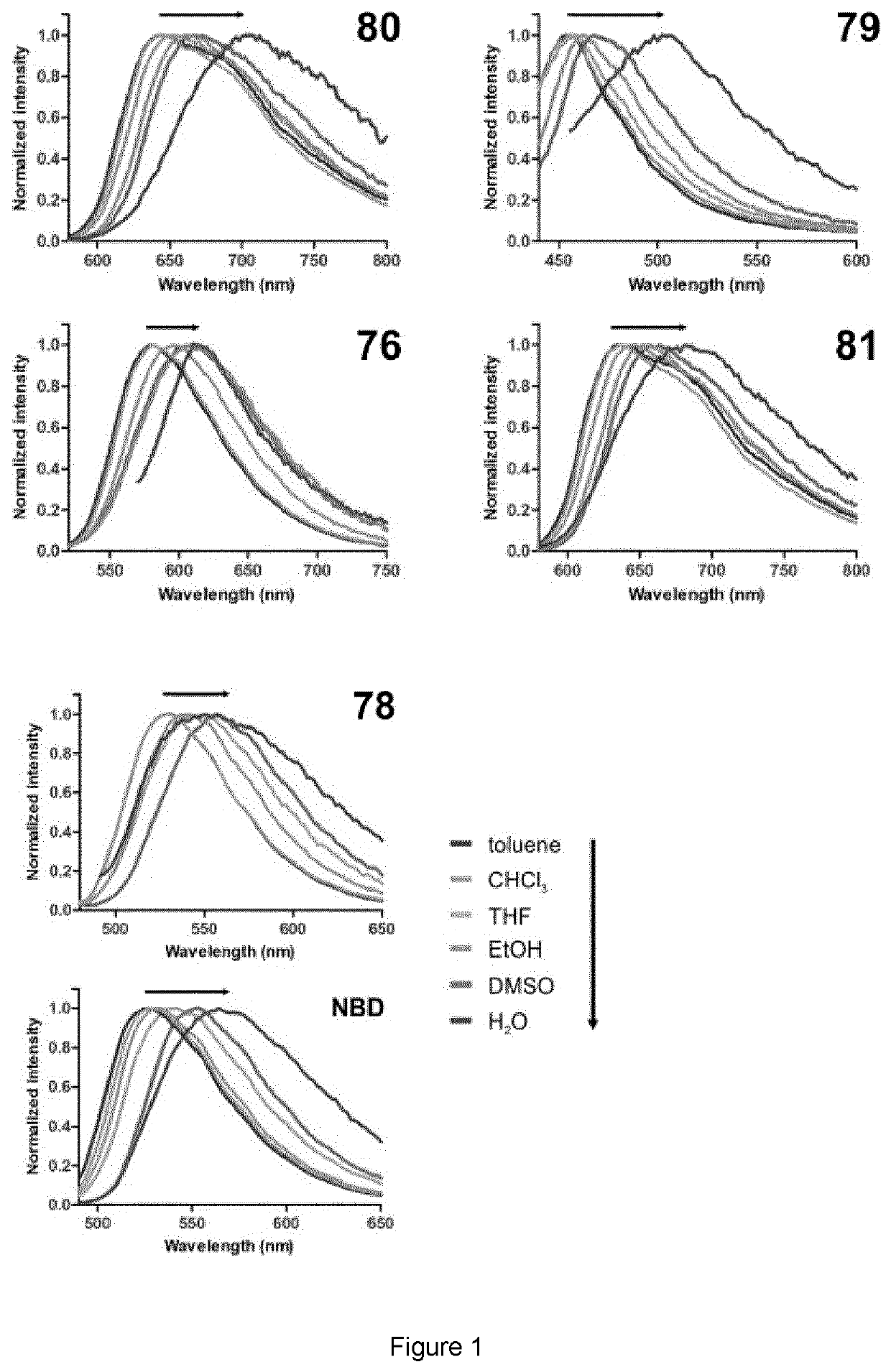
However, NBD (emission˜540 nm) is incompatible with other green fluorescent reporters (e.g. GFP) and has limited application for in vivo use.

Method used


Image

  • Small tunable fluorophores for the detection and imaging of biomolecules
  • Small tunable fluorophores for the detection and imaging of biomolecules
  • Small tunable fluorophores for the detection and imaging of biomolecules

Examples


Embodiment Construction

[0068]The preparation of SCOTfluors was achieved in two synthetic steps from a common intermediate of formula II. The detailed preparation of the Scotfluors is described below as well as the analytical methods used in the examples.

[0069]General Materials

[0070]Commercially available reagents were used without further purification. Thin-layer chromatography was conducted on Merck silica gel 60 F254 sheets and visualized by UV (254 and 365 nm). Silica gel (particle size 35-70 μm) was used for column chromatography. 1H and 13C spectra were recorded in a Bruker Avance 500 spectrometer (at 500 and 126 MHz, respectively). Data for 1H NMR spectra are reported as chemical shift δ (ppm), multiplicity, coupling constant (Hz), and integration. Data for 13C NMR spectra are reported as chemical shifts relative to the solvent peak. HPLC-MS analysis was performed on a Waters Alliance 2695 separation module connected to a Waters PDA2996 photo-diode array detector and a ZQ Micromass mass spectrometer...


PUM

PropertyMeasurementUnit
Time-aaaaaaaaaa
particle sizeaaaaaaaaaa
pHaaaaaaaaaa

Abstract

The invention relates to small, conjugatable, orthogonal and tunable fluorophores for imaging of small bioactive molecules. The invention further relates to processes for the preparation of the compounds, and uses of the compounds in therapeutic, diagnostic, surgery and analytical applications. The invention provides a compound of formula (I), a derivative or a salt thereof. Wherein X is selected from the group consisting of NH, O, S, SeR5R6, CR7R8; R1 is selected from the group consisting of amines, alcohols, thiols, thiophenols, selenols, selenophenols and aryl groups; R2 and R3 are independently H or a halogen; R4 tis either H, nitro or cyano; R5 is either absent or methyl or oxygen; R6 is either absent or methyl or oxygen; and R7 and R8 are independently selected from the group consisting of linear or cyclic alkyl groups containing halogen, amino, cyano or carboxylic ester substituents, and alkyl aryl groups.

Description

FIELD OF THE INVENTION[0001]The invention relates to small, conjugatable, orthogonal and tunable fluorophores for imaging of small bioactive molecules. The invention further relates to processes for the preparation of the compounds, and uses of the compounds in therapeutic, diagnostic, surgery and analytical applications.BACKGROUND OF THE INVENTION[0002]Metabolites are essential biochemical components, with their transport and localization regulating most biological functions. Despite advances in fluorescence imaging to label small biomolecules, there are few approaches to image in situ small metabolites in live cells and intact organisms.[0003]Most metabolites do not contain groups that allow direct visualization and need to be modified with exogenous chromophores. Most in vivo imaging fluorophores rely on near-infrared (NIR) chemical scaffolds as they allow deep penetration with minimal photodamage and low tissue autofluorescence. However, fluorescent labels, in particular red and...

Claims


Application Information

Patent Timeline
Patent Type & Authority Applications(United States)
IPC IPC(8): C09B51/00C07D249/18A61K49/00C07D285/14C07D293/12C07D235/08A61B5/00A61B3/12G01N21/64
CPCC09B51/00C07D249/18A61K49/0021C07D285/14C07D293/12G01N2021/6439A61B5/0066A61B5/0071A61B5/0095A61B3/1241G01N21/6428C07D235/08C09B23/04C09B23/0058C07D293/10C07D403/04C07D421/12C07D417/12C07D405/12C07D235/00C07D235/18C07D421/04C07D417/04C07D235/16C09K11/06C09K2211/1055C09K2211/1007C09K2211/1051C09K2211/1059C09K2211/1044C09K2211/1029C09K2211/1088C09B57/00A61B5/0075
Inventor VENDRELL, MARCFERNANDEZ, ANTONIOBENSON, SAMBARTH, NICOLE D.DE MOLINER, FABIO
Owner THE UNIV COURT OF THE UNIV OF EDINBURGH
Learn More