
High-content imaging of microfluidic devices

Pending Publication Date: 2021-11-04
EMULATE INC
View PDF0 Cites 0 Cited by
  • Summary
  • Abstract
  • Description
  • Claims
  • Application Information

AI Technical Summary

Benefits of technology

The present invention is related to a method and system for high-speed and high-quality microscopy imaging of microfluidic cell culture systems. This invention aims to reduce the variability in imaging results and improve the accuracy of analysis. It includes a statistical analysis method and a special adaptor designed to improve the image quality of microfluidic devices. Overall, this invention provides a more reliable and efficient tool for studying cell cultures and their interactions in a microfluidic environment.

Problems solved by technology

The relative simplicity of two-dimensional cell culture models means that findings often do not translate into the clinical phases of drug development.
However, gene therapy treatment has risks including undesirable inflammation, e.g. causing tissue damage, causing organ damage leading to organ failure; targeting healthy cells causing damage that may lead to other illness, diseases and cancer; gene therapy vectors themselves may recover their ability to cause disease and do so; genetic material is inserted into an unintended location with a host cell gene which may lead to tumor formation or other unwanted consequence.

Method used


Image

  • High-content imaging of microfluidic devices
  • High-content imaging of microfluidic devices
  • High-content imaging of microfluidic devices

Examples


Embodiment Construction

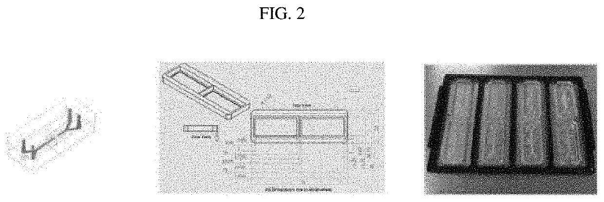
culture systems. A method of high-content microfluidic device microscopy is contemplated, along with related statistical analysis and microfluidic device adaptors.

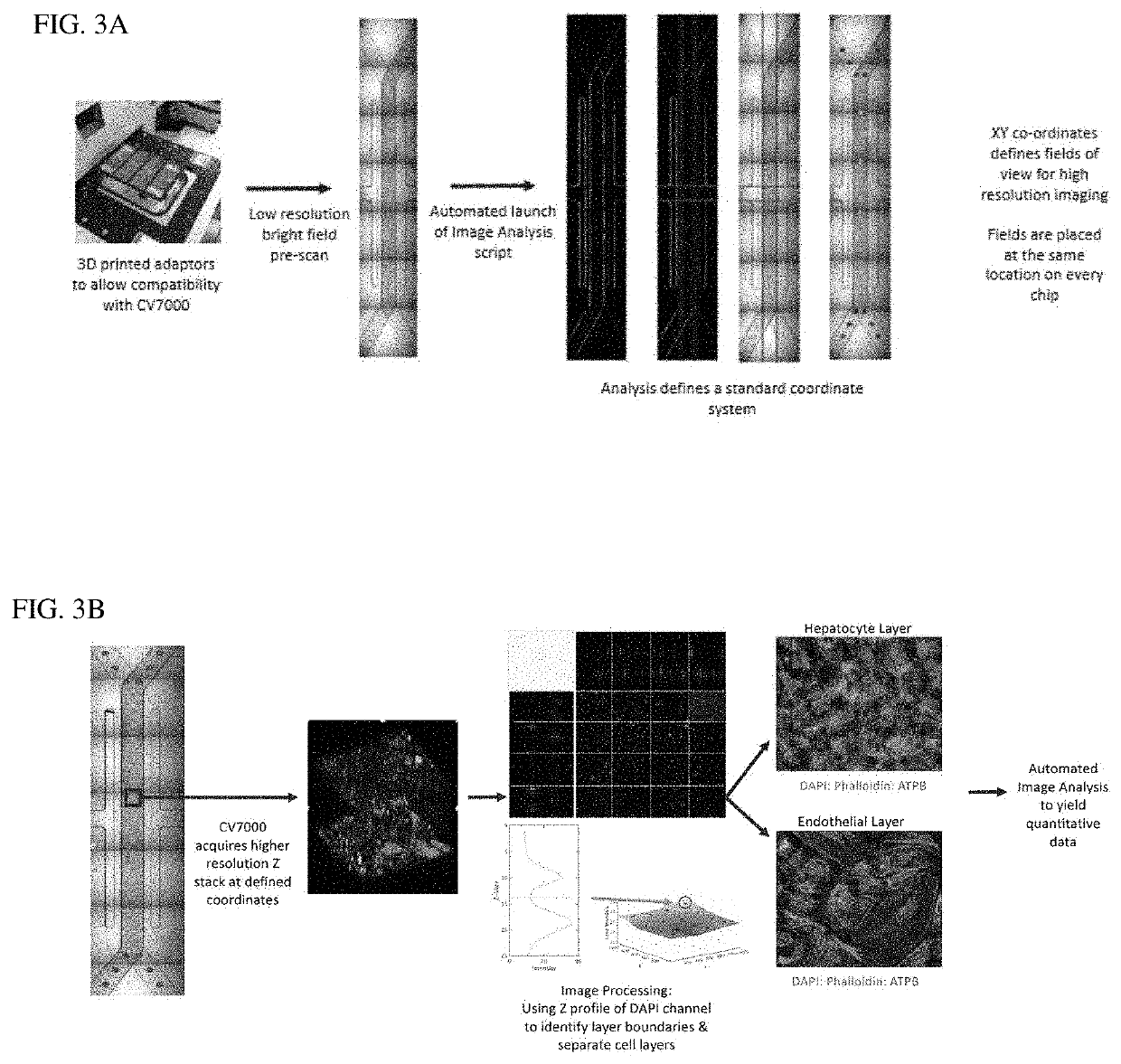
[0091]Described herein is a novel end-to-end, automated workflow to capture and analyze confocal images of microfluidic devices containing multi-cellular organ culture in order to assess detailed cellular phenotype across large batches of microfluidic devices. By automating this process, not only is acquisition time reduced, but process variability and user bias is also minimized. Automation has enabled establishment, for the first time, of a framework of statistical best practice for microfluidic device imaging, creating the capability of using microfluidic devices and imaging for routine testing in drug discovery applications that rely on quantitative image data for decision making. The approach was tested using test compounds, such as compounds whose mechanism of toxicity was linked to mitochondrial damage with subseque...


PUM


Abstract

The present invention is related to high-content microscopy imaging of microfluidic cell culture systems. A method of high-content microfluidic device microscopy is contemplated. along with related statistical analysis and microfluidic device adaptors.

Description

CROSS-REFERENCE TO RELATED APPLICATIONS[0001]This application claims priority to U.S. Provisional Patent Applications No. 62 / 795,345 filed Jan. 22, 2019; 62 / 828,849 filed Apr. 3, 2019; 62 / 834,004 filed Apr. 15, 2019; and Ser. No. 37 / 487,696 filed Oct. 17, 2019, herein incorporated by reference in their entireties.FIELD OF INVENTION[0002]The present invention is related to high-content microscopy imaging of microfluidic cell culture systems. A method of high-content microfluidic device microscopy is contemplated, along with related statistical analysis and microfluidic device adaptors.BACKGROUND[0003]The pharmaceutical industry needs to improve the probability of success of drugs reaching late stage clinical trial. One category of microfluidic devices are micro-engineered systems that aim to recapitulate the organ microenvironment for drug discovery. Microfluidic devices comprising cells of human origin have been adopted for enhancing pre-clinical efficacy and toxicity evaluation and...

Claims


Application Information

Patent Timeline
IPC IPC(8): G01N15/14G01N33/50G02B21/00
CPCG01N15/1434G01N33/5067G01N2015/144G02B21/0072G02B21/008G02B21/34G02B21/24G01N33/5044G06V20/693G01N15/1484G01N2015/1493G01N2015/1497G01N2015/1006G01N15/0227G01N15/01G01N15/1433G01N15/14G01N33/48G02B21/02C12N15/8645C12N2750/14143G01N33/5008G01N2500/10C12N15/86C12N15/88C12Q1/02
Inventor JANG, KYUNG-JINLEVNER, DANIELKODELLA, KONSTANTIA-ROUMVINIRUBINS, JONATHANPETROPOLIS, DEBORA BARREIROSPEEL, SAMATHACORRIGAN, ADAM M.EHRARDT, BEATEPINTO, PEDROWILLIAMS, DOMINICBOECKELER, MATTFOSTER, ALISON J.HAMILTON, GERALDINEEWART, LORNA EWART
Owner EMULATE INC
Learn More