Eureka AIR delivers breakthrough ideas for toughest innovation challenges, trusted by R&D personnel around the world.

Neural Network Classification of Osteolysis and Synovitis Near Metal Implants

a neural network and osteolysis technology, applied in the field of neural network classification of osteolysis and synovitis near metal implants, can solve the problems of failures requiring revision, complex mri interpretation in the immediate vicinity, and poor characterization of clinically relevant mri signatures

Pending Publication Date: 2020-12-31
THE MEDICAL COLLEGE OF WISCONSIN INC
View PDF4 Cites 3 Cited by
  • Summary
  • Abstract
  • Description
  • Claims
  • Application Information

AI Technical Summary

Benefits of technology

The patent text describes a method for generating a feature map that depicts a spatial distribution or pattern of a feature associated with osteolysis or synovitis. This is done by accessing magnetic resonance imaging (MRI) data from a subject and inputting it into a trained machine learning algorithm that has been trained on training data. The generated output includes at least one feature map that depicts a spatial distribution of the feature associated with osteolysis or synovitis. The patent also describes a method for constructing and implementing a machine learning algorithm to a feature map that depicts spatial patterns of a feature associated with osteolysis or synovitis across a region in a subject. Additionally, the patent describes a method for generating feature data that indicates a thickness of a synovial membrane in a subject by accessing MRI data and inputting it into a trained machine learning algorithm. The technical effects of the patent text include improved diagnostic accuracy for identifying and characterizing joint disorders associated with osteolysis or synovitis.

Problems solved by technology

In practice, most HA failures requiring revision are due to alterations in adjoining tissues (e.g., bone, synovium, muscle).
However, the mitigation of the physical challenge of MRI metal artifacts has exposed another challenge: the complexity of MRI interpretation in the immediate vicinity of HA.
This challenge, combined with the complex manifestation of the MRI findings, results in poor characterization of these clinically relevant MRI signatures and an under-utilization of the imaging technology in many clinical orthopedic imaging environments.

Method used


Image

  • Neural Network Classification of Osteolysis and Synovitis Near Metal Implants
  • Neural Network Classification of Osteolysis and Synovitis Near Metal Implants
  • Neural Network Classification of Osteolysis and Synovitis Near Metal Implants

Examples


Embodiment Construction

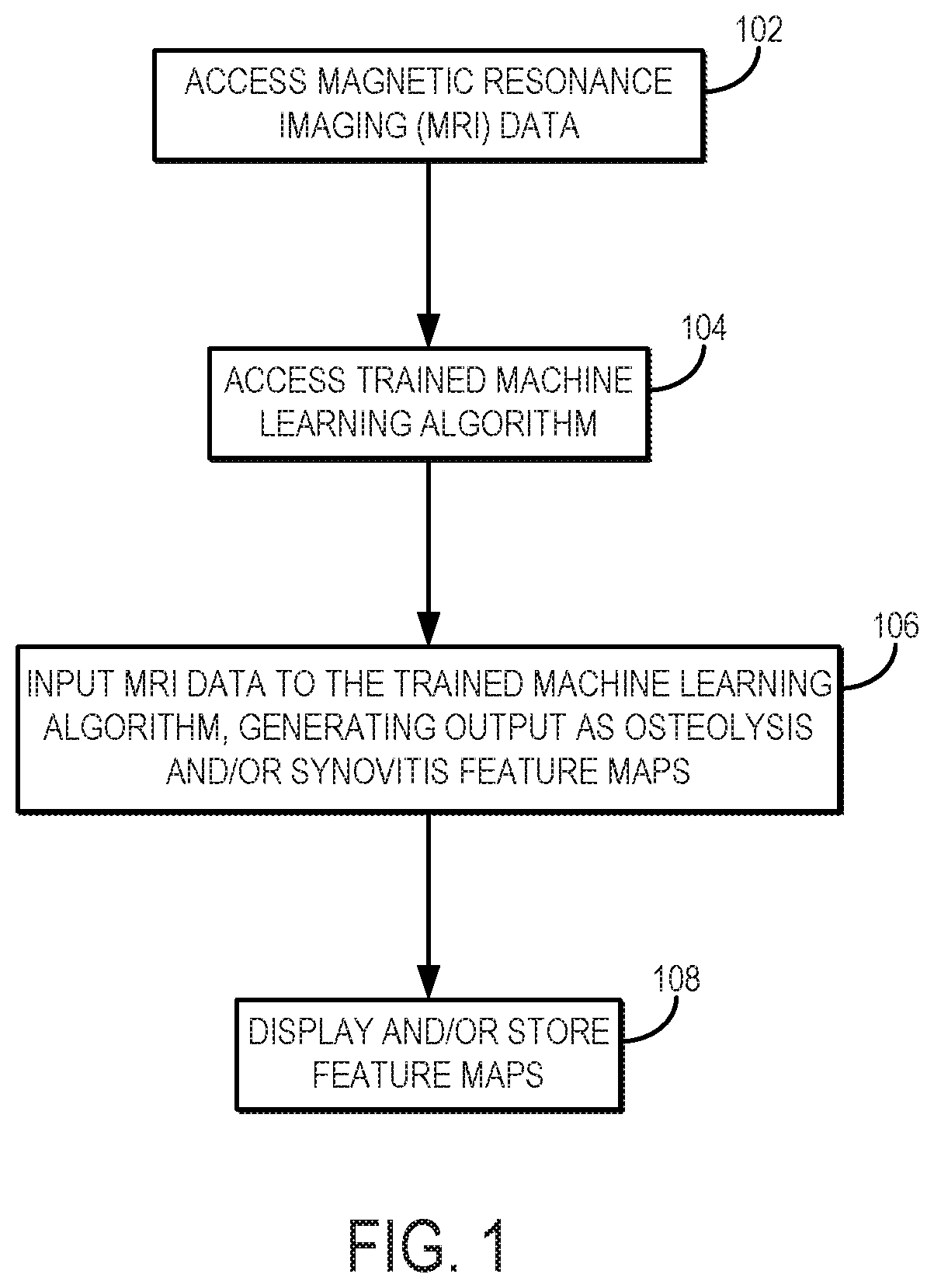
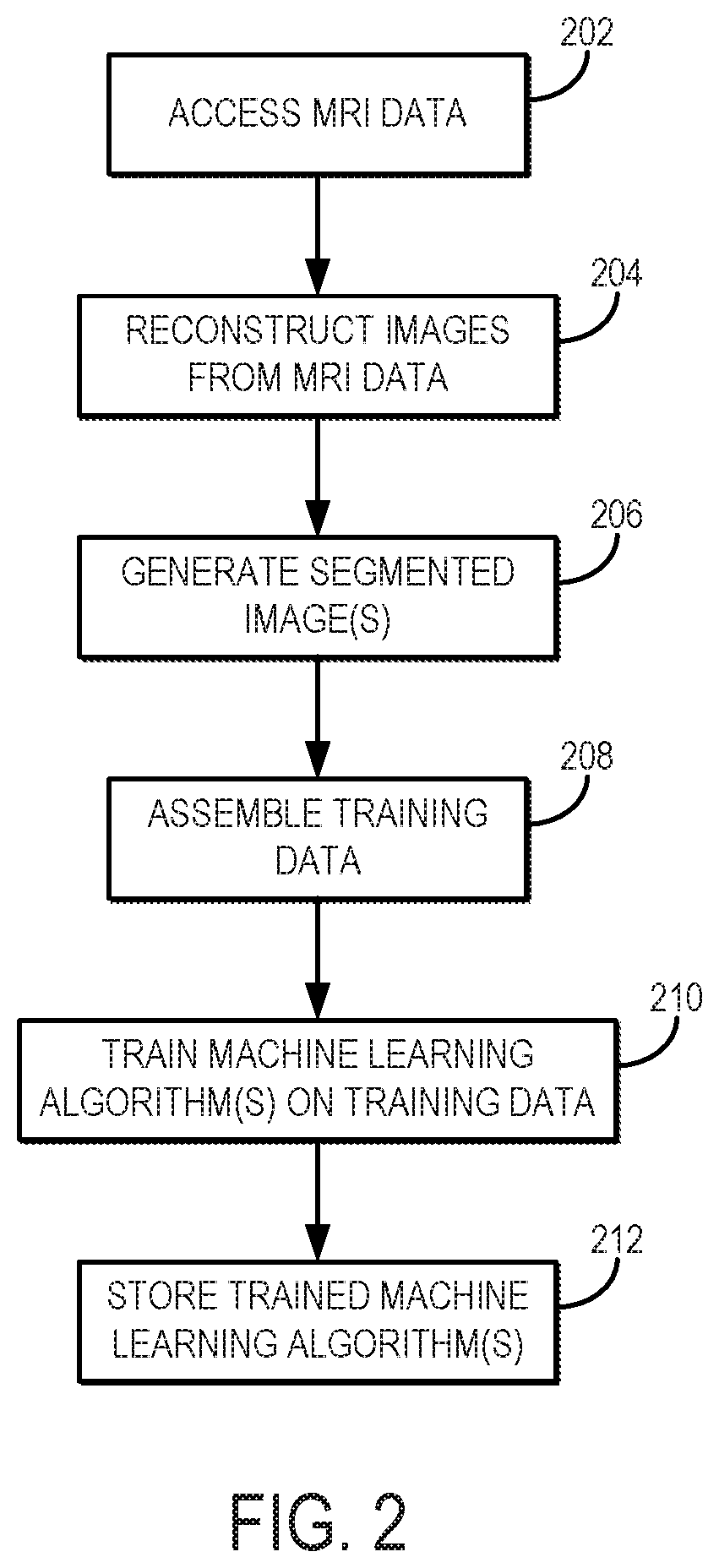
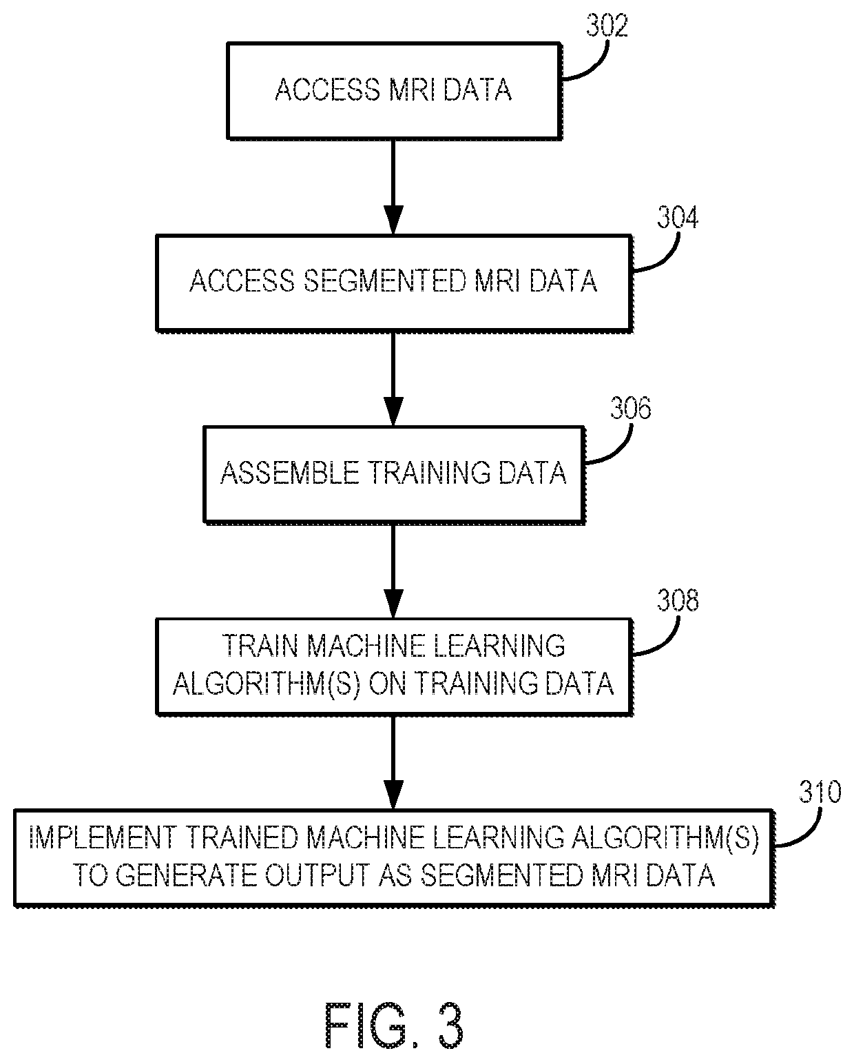
[0017]Described here are systems and methods for utilizing a machine learning algorithm implemented with a hardware processor and memory to provide image-based deep learning on 3D-MSI MRI to provide supplementary guidance for radiological identification of common soft-tissue pathologies near HA or other metallic implants or objects. In particular, preliminary data demonstrates the promising capabilities of convolutional neural networks (“CNNs”) trained on a cohort of normal and pathological 3D-MSI of HA (or patients with or other metallic implants or objects) to spatially identify periprosthetic bone loss (i.e., osteolysis) and adverse synovial responses. Using this artificial intelligence (“AI”) analysis engine, feature maps advising potential soft tissue and bone pathology can be constructed on a subject-specific basis and presented to radiologists and surgeons as a direct visual presentation of regional tissue abnormalities near HA or other metallic implants or objects. This comp...


PUM


Abstract

Systems and methods for training and implementing a machine learning algorithm to generate feature maps depicting spatial patterns of features associated with osteolysis, synovitis, or both. MRI data, including multispectral imaging data, are input to the trained machine learning algorithm to generate the feature maps, which may indicate features such as a location and probability of a pathology classification, a severity of synovitis, a type of synovitis, a synovial membrane thickness, and other features associated with osteolysis or synovitis. In some implementations, synovial anatomy are segmented in the MRI data before inputting the MRI data to the machine learning algorithm. These segmented MRI data may be generated using another trained machine learning algorithm.

Description

CROSS-REFERENCE TO RELATED APPLICATIONS[0001]This application claims the benefit of U.S. Provisional Patent Application Ser. No. 62 / 637,812, filed on Mar. 2, 2018, and entitled “NEURAL NETWORK CLASSIFICATION OF OSTEOLYSIS AND SYNOVITIS NEAR METAL IMPLANTS,” which is herein incorporated by reference in its entirety.STATEMENT REGARDING FEDERALLY SPONSORED RESEARCH[0002]This invention was made with government support under AR064840 awarded by the National Instituted of Health. The government has certain rights in the invention.BACKGROUND[0003]An estimated 2.5 million Americans currently have artificial hip joints, or hip arthroplasty (“HA”), all of which have inevitable failure modes that depend heavily on patient-specific risk and genetic factors. In practice, most HA failures requiring revision are due to alterations in adjoining tissues (e.g., bone, synovium, muscle). These tissue alterations are best assessed using magnetic resonance imaging (“MRI”). In particular, MRI can visualiz...

Claims


Application Information

Patent Timeline
IPC IPC(8): G06T7/00G06N3/08G06N3/04G16H30/20G16H50/20G16H30/40G16H50/50G16H50/70G16H50/30G06T7/11G06K9/62A61B5/055A61B5/00
CPCG16H50/20G06T7/0012G06N3/088G16H50/70G06T2207/10088A61B2576/02G06K9/6267G16H30/20G06T7/11G06T2207/20081A61B5/4504A61B5/4851G06K9/6232G06T2207/30008A61B5/7267G06N3/0454G16H50/50A61B5/055G06T2207/20084G16H30/40G16H50/30G01R33/5608G06F18/24G06N3/045
Inventor KOCH, KEVIN M.NENCKA, ANDREW S.KARR, ROBIN A.SWEARINGEN, BRADLEY J.POTTER, HOLLISKOFF, MATTHEW F.
Owner THE MEDICAL COLLEGE OF WISCONSIN INC
Why Eureka
Eureka Blog
Learn More