
Methods for diagnosing and treating prostate cancer

a prostate cancer and prostate cancer technology, applied in the direction of lysates, instruments, drug compositions, etc., can solve the problems of poor specificity of psa test, patient discomfor

Inactive Publication Date: 2020-08-06
CORNELL UNIVERSITY
View PDF0 Cites 0 Cited by
  • Summary
  • Abstract
  • Description
  • Claims
  • Application Information

AI Technical Summary

Benefits of technology

This approach effectively inhibits prostate cancer cell proliferation, provides a non-invasive diagnostic tool for prostate cancer, and aids in selecting treatment options based on sAC expression levels, improving the specificity and accuracy of prostate cancer diagnosis and treatment.

Problems solved by technology

While the PSA test has resulted in the majority of prostate cancer cases being diagnosed in asymptomatic men (Mettlin et al., Cancer, 83(8): 1679-1684 (1998a); Mettlin et al., Cancer, 82(2): 249-251 (1998b); Humphrey et al., J. Urol., 155: 816-820 (1996); and Grossfeld et al., Epidemiol. Rev., 23(1): 173-180 (2001)), the PSA test suffers from poor specificity, which can be as low as 33% when a PSA cut-off level of 2.6 ng / mL is used (Thompson et al., N. Engl. J. Med., 350: 2239-2246 (2004)), even though the sensitivity can be as high as 83%.
The poor specificity of the PSA test is a direct result of increased secretion of PSA in other diseases of the prostate, such as benign prostate hyperplasia (BPH) and prostatitis.
Over 1 million needle biopsies of prostates are performed each year at a cost of about $1,500 each and much discomfort to the patient.
However, less than 200,000 of these result in a diagnosis of prostate cancer.
These markers, however, lack the specificity needed for consistently reliable diagnoses.
Similarly, prognostic biomarkers such as the TMPRSS2-ERG gene fusion, PTEN deletion, and SPINK1 overexpression also lack the specificity to assess a wide range of prostate cancers, leaving a significant number of prostate cancers without further prognostic information apart from calculating a cancer's Gleason score.
Currently there are no known biomarkers that can indicate prostate cancers that have invaded into the periprostatic soft tissue.

Method used


Image

  • Methods for diagnosing and treating prostate cancer
  • Methods for diagnosing and treating prostate cancer
  • Methods for diagnosing and treating prostate cancer

Examples


example 1

[0044]This example demonstrates that sAC protein is overproduced in prostate cancer cells.

[0045]The sAC gene expression, sAC protein subcellular localization, and sAC protein distribution in both benign and malignant prostate tissue samples were examined. Immunohistochemical staining using a mouse monoclonal sAC antibody (R21) was performed on both tumor and benign tissue from 12 radical prostatectomy specimens. Two tumors were well-differentiated (Gleason score 6), 7 were moderately differentiated (Gleason score 7), and 3 were poorly differentiated (Gleason score 8-10).

[0046]Briefly, five micron-thick sections of the formalin-fixed paraffin-embedded tissue were deparaffinized and stained using a Bond III Autostainer (Leica Microsystems, Buffalo Grove, Ill.) and the manufacturer's Heat-Induced Epitope Retrieval 1 protocol with supplied reagents. Mouse monoclonal R21 sAC antibody (CEPBiotech, Inc, R21-IHC, Tamarac, Fla.) was used at a dilution of 1:750 as previously described (Zippin...

example 2

[0052]This example demonstrates that sAC protein is overproduced in prostate cancer cells.

[0053]Prostate cancer cases were retrospectively identified from the database of the Division of Surgical Pathology, Weill Cornell Medical College. Tissue microarrays (TMAs) were constructed from the archival formalin-fixed, paraffin-embedded tissue samples using 0.6 mm cores, with each area represented in triplicate. When possible, areas of benign prostatic tissue, high grade prostatic intraepithelial neoplasia (HGPIN), and invasive prostatic adenocarcinoma were all sampled from each case; however, in some cases not all tissue types were present for sampling / evaluation.

[0054]Immunohistochemical staining for three sAC antibodies (R21, R40, and R52) was performed on the TMA slides. The TMAs included 50 samples of benign prostatic tissue, 35 samples of HGPIN, 65 samples of localized prostatic adenocarcinoma, and 25 samples of neuroendocrine prostate cancer (NEPC), castration resistant prostate ca...

example 3

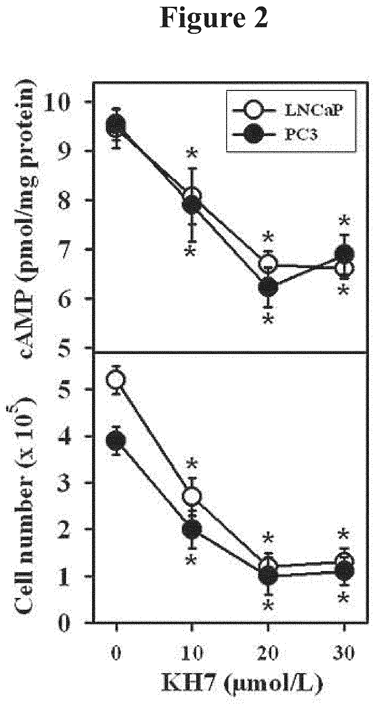
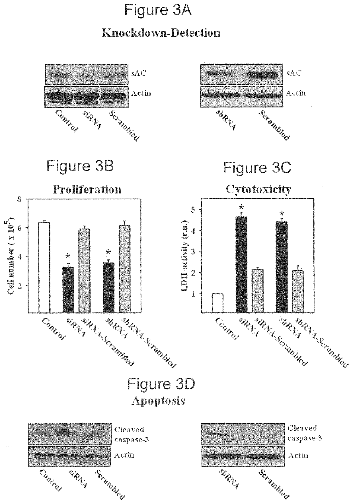
[0061]This example demonstrates a method of inhibiting proliferation of prostate cancer cells by suppressing the activity of soluble adenylyl cyclase (sAC) protein.

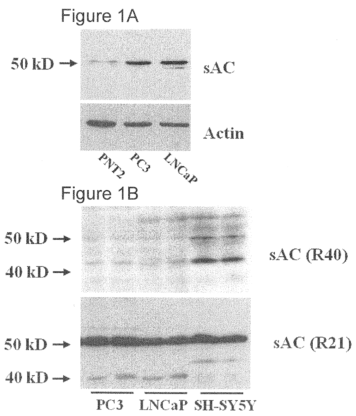
[0062]The androgen-sensitive LNCaP (ATCC-Nr. CRL-1740D) human prostate carcinoma cell line, the androgen-insensitive PC3 (ATCC-Nr. CRL-1435D) human prostate carcinoma cell line, and neuroblastoma cell line SH-SY5Y (ATCC-Nr. CRL-2266) were purchased from the American Type Culture Collection, and human normal prostate epithelial cell line PNT2 was purchased from Sigma-Aldrich (Cat. Nr. 95012613). Cells were expanded and frozen in aliquots within four weeks of purchase. Cells were thawed and cultured for no more than three further passages. PNT2 cells were cultured in medium RPMI1640 supplemented with 10% fetal calf serum, glutamine and antibiotics. All other cells were cultured in Dulbecco's modified Eagle's medium that was supplemented with 5% fetal calf serum, glutamine, and antibiotics. The cells (1.5×10⁵) were seeded in...


PUM


Abstract

The invention is directed to a method of inhibiting prostate cancer cell proliferation using a substance that inhibits the activity of soluble adenylyl cyclase (sAC) protein. The invention also is directed to methods of diagnosing and prognosticating prostate cancer in a subject by evaluating sAC gene expression or sAC protein production in the subject.

Description

STATEMENT REGARDING FEDERALLY SPONSORED RESEARCH AND DEVELOPMENT[0001]This invention was made with Government support under Grant Number NCI K08 CA 160657 awarded by the National Cancer Institute (NCI). The Government has certain rights in this invention.INCORPORATION-BY-REFERENCE OF MATERIAL SUBMITTED ELECTRONICALLY[0002]Incorporated by reference in its entirety herein is a computer-readable nucleotide / amino acid sequence listing submitted concurrently herewith and identified as follows: One 4,225 Byte ASCII (Text) file named “715739_ST25.TXT,” created on Dec. 11, 2013.BACKGROUND OF THE INVENTION[0003]Prostate cancer is the most common malignancy and the second leading cause of death among men in the U.S. (Li et al., Biochim. Biophys. Acta, 1704: 87-102 (2004)). The National Cancer Institute (NCI) estimates that in 2013, over 230,000 new cases of prostate cancer will be diagnosed, and over 29,000 men will die of prostate cancer in the United States. The prostate-specific antigen or...

Claims


Application Information

Patent Timeline
Patent Type & Authority Applications(United States)
IPC IPC(8): A61K31/4184A61K31/713A61K31/7105G01N33/574C12N15/113
CPCC12N2310/14A61K31/713C12N2310/531C12N15/1137C12N2320/30G01N2333/988C12Y406/01001G01N33/57434A61K31/7105A61K31/4184A61P35/00C12N15/11
Inventor ROBINSON, BRIANZIPPIN, JONATHAN
Owner CORNELL UNIVERSITY
Learn More