
Compositions and Methods for Treating a Disorder or Defect in Soft Tissue

a technology of soft tissue and compositions, applied in the direction of drug compositions, dermatological disorders, cardiovascular disorders, etc., can solve the problems of increasing the risk of medical complications, intense pain and restricted movement, and limited success of long-term correction of structures in the body

Inactive Publication Date: 2019-05-09
DREXEL UNIV
View PDF0 Cites 1 Cited by
  • Summary
  • Abstract
  • Description
  • Claims
  • Application Information

AI Technical Summary

Benefits of technology

[0266]An advantage of the present invention is that the entire intervertebral disc is not removed in order to effect treatment of the degenerated disc. However, it is recognized that in some instances, the materials of the present invention can be administered into the degenerated disc without removing native material from the degenerated disc prior to administration of the materials. The purpose of removing native material from the degenerated disc is to make room for the materials to be administered.
[0267]When cells are used to treat a degenerated disc, the cells may be administered to a mammal following a period of in vitro culturing. The cell may be cultured in a manner that induces the cell to differentiate in vitro. However, the cells can be administered into the recipient in an undifferentiated state where the administered cells differentiate to express at least one characteristic of a disc cell in vivo in the mammal.
[0268]The cells of this invention can be transplanted into a mammal using techniques known in the art such as i.e., those described in U.S. Pat. No 5,618,531, which is incorporated herein by reference, or into any other suitable site in the body. Transplantation of the cells of the present invention can be accomplished using techniques well known in the art as well as those described herein, or using techniques developed in the future. The present invention comprises a method for transplanting, grafting, infusing, or otherwise introducing the cells into a mammal, preferably, a human.
[0269]The cells can be suspended in an appropriate diluents. Suitable excipients for administration solutions are those that are biologically and physiologically compatible with the cells and with the recipient, such as buffered saline solution or other suitable excipients. The composition for administration can be formulated, produced and stored according to standard methods complying with proper sterility and stability.
[0270]The cells may also be encapsulated and used to deliver biologically active molecules, according to known encapsulation technologies, including microencapsulation (see, e.g., U.S. Pat Nos. 4,352,883; 4,353,888; and 5,084,350, herein incorporated by reference), or macroencapsulation (see, e.g., U.S. Pat. Nos. 5,284,761; 5,158,881; 4,976,859; and 4,968,733; and International Publication Nos. WO 92 / 19195; WO 95 / 05452, all of which are incorporated herein by reference). For macroencapsulation, the number of cells used in the devices can be varied. Several macroencapsulation devices may be administered in the mammal. Methods for macroencapsulation and administration of cells are well known in the art and are described in, for example, U.S. Pat. No. 6,498,018.
[0271]The mode of administration of the cells of the invention to the mammal may vary depending on several factors including the type of disease being treated, the age of the mammal, whether the cells are differentiated or not, whether the cells have exogenous DNA introduced therein, and the like. The cells may be introduced to the desired site by direct administration, or by any other means used in the art for the introduction of compounds administered to a mammal suffering from a particular disease or disorder of the disc.

Problems solved by technology

However, these approaches have met with limited success for the long-term correction of structures in the body.
Degenerated and damaged soft tissues of the musculoskeletal system cause and increase the risk of medical complications resulting in intense pain and restricted motion.
Soft tissue degeneration of the ligaments and intervertebral discs also increase the risk of damage to and back pain from local spinal joints, including: zygapophysical (facet), costovertebral, sacroiliac, sacral vertebral and atlantoaxial joints.
The repair of large segmental defects in diaphyseal bone is a significant problem faced by orthopaedic surgeons today.
The use of fresh autologous bone graft material has been viewed as the historical standard of treatment but is associated with substantial morbidity including infection, malformation, pain, and loss of function (Kahn et al., 1995, Clin. Orthop. Rel. Res. 313:69-75).
Although widely used for many years, the risk of disease transmission, host rejection, and lack of osteoinduction compromise its desirability (Leads, 1988, JAMA 260:2487-2488).
The favorably porous nature of these implants facilitate bony ingrowth, but their lack of osteoinductive potential limits their utility.
Though sound in principle, the practicality of obtaining enough bone marrow with the requisite number of osteoprogenitor cells is limiting.
The leading cause of back pain is due to degeneration of the intervertebral disc.
This degeneration leads to additional changes in the spine as the disc degenerates and loses height.
This reduces the ability of the disc to share load, which in turn causes the annulus to carry more load.
This causes the annulus to degenerate.
Artificial discs are challenged by both biological and biomechanical considerations, and often require complex prosthesis designs.
One limitation of the nucleus replacement procedure resides in the need of relatively intact annulus and endplates, which means the nucleus replacement procedure must be performed when disc degeneration is at an early stage.
In many instances, for example, when these devices are implanted in the body, they are subject to a “foreign body” response from the surrounding host tissues.
Encapsulation of surgical implants complicates a variety of reconstructive and cosmetic surgeries, and is particularly problematic in the case of breast reconstruction surgery where the breast implant becomes encapsulated by a fibrous connective tissue capsule that alters the anatomy and function.
Capsular (fibrous) contractures can result in hardening of the breast, loss of the normal anatomy and contour of the breast, discomfort, weakening and rupture of the implant shell, asymmetry, infection, and patient dissatisfaction.
Further, fibrous encapsulation of any soft tissue implant can occur even after a successful implantation if the device is manipulated or irritated by the daily activities of the patient.
For example, unwanted scarring can result from surgical trauma to the anatomical structures and tissue surrounding the implant during the implantation of the device.
Bleeding in and around the implant can also trigger a biological cascade that ultimately leads to excess scar tissue formation.
Similarly, if the implant initiates a foreign body response, the surrounding tissue can be inadvertently damaged from the resulting inflammation, leading to loss of function, tissue damage and / or tissue necrosis.
Furthermore, certain types of implantable prostheses (such as breast implants) include gel fillers (e.g., silicone) that tend to leak through the membrane envelope of the implant and can potentially cause a chronic inflammatory response in the surrounding tissue (which augments tissue encapsulation and contracture formation).
When scarring occurs around the implanted device, the characteristics of the implant-tissue interface degrade, the subcutaneous tissue can harden and contract and the device can become disfigured.
The effects of unwanted scarring in the vicinity of the implant are the leading cause of additional surgeries to correct defects, break down scar tissue, or remove the implant.

Method used


Image

  • Compositions and Methods for Treating a Disorder or Defect in Soft Tissue
  • Compositions and Methods for Treating a Disorder or Defect in Soft Tissue
  • Compositions and Methods for Treating a Disorder or Defect in Soft Tissue

Examples


example 1

Effect of Aging and Degeneration on Fluid exchange, Stress Concentrations and Osmotic Pressure of the Human Intervertebral Disc During the Diurnal Cycle

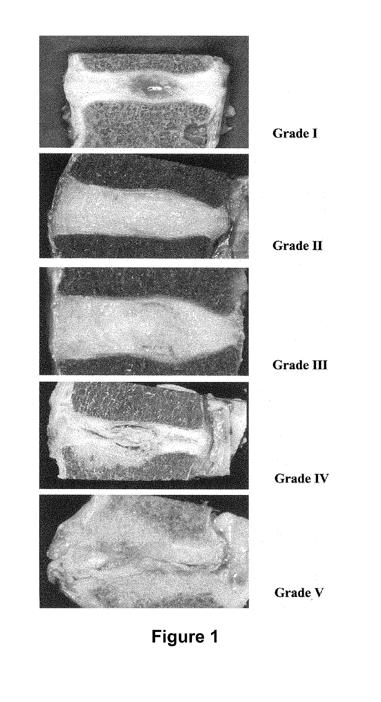
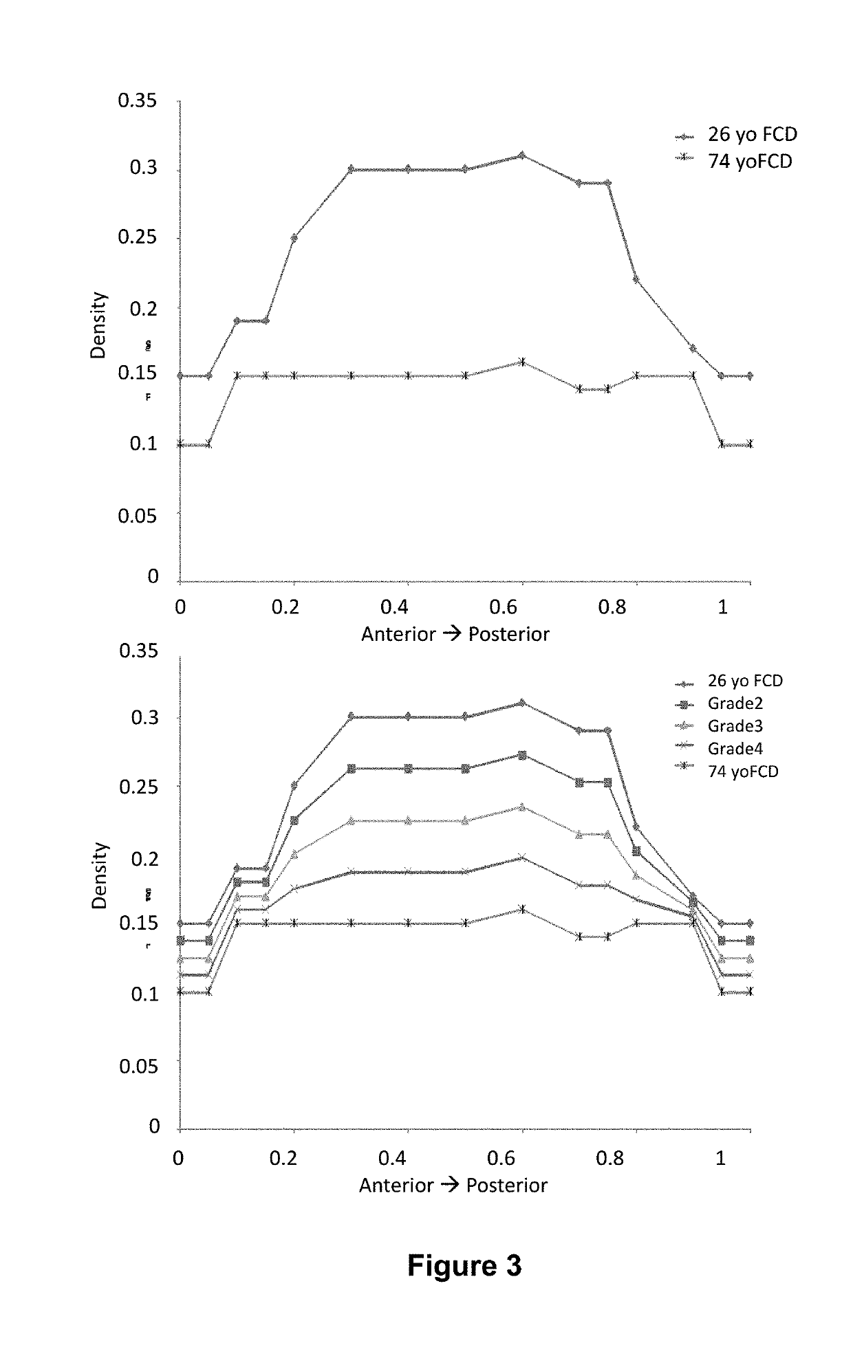
[0315]The human intervertebral disc is the primary compression-carrying component of the spine. Its roles are to transmit and distribute loads, and allow for the necessary flexibility of the spine. It is comprised of a central gel-like nucleus pulposus, an outer annulus fibrosus, and upper and lower endplates consisting of cartilaginous and bony portions. During a diurnal cycle, the intervertebral disc experiences approximately 16 hours of functional loading (standing, sitting, etc.), followed by 8 hours of recovery (lying prone). Therefore, the fluid lost during the loading period must be replenished in half the time. As the disc is compressed and fluid is exuded, the density of the fixed charges within the nucleus pulposus is increased, creating an osmotic gradient with the interstitial fluid surrounding the disc. This osmotic pote...

example 2

Restoration of Proteoglycan to the Nucleus Pulposus of the Intervertebral Disc

[0331]The intervertebral disc is the largest avascular tissue in the human body and is mainly comprised of three different tissues. The central core, the nucleus pulposus, is surrounded by the outer annulus fibrosus and the upper and lower cartilaginous endplates. Lower back pain was reported in more than 80% of the cases exhibiting degeneration of lumbar intervertebral discs (Luoma et al., 2000, Spine 25(4):487). With aging, the proteoglycan and water content in the central nucleus reduces significantly, causing abnormal loading to the outer annulus (Urban et al., 1988, Spine (Phila Pa 1976) 13(2):179-87; Luoma et al., 2000, Spine 25(4):487; Yerramalli et al., 2007, Biomechanics and Modeling in Mechanobiology 6(1):13-20; Urban et al., 2003, Arthritis Research and Therapy 5(3):120-38; Roughley et al., 2002, Biochemical Society Transactions. 30:869-74; Tropiano et al., 2005, The Journal of Bone and Joint Su...

example 3

Nucleus Pulposus Augmentation

[0343]Prior work has investigated the role of the nucleus pulposus in human lumbar intervertebral disc mechanics. The nucleus is critical to the stability of the disc through the neutral zone (Joshi et al., 2008, J Biomech. 41(10):2014-111). After denucleation of the intervertebral disc, the neutral zone as well as the full range of motion was shown to increase significantly over the same measurements for the intact disc to which they were normalized. In addition, the stiffness of the disc through the neutral zone region was significantly reduced from that of the intact disc. This study shows that the nucleus is critical in providing stability to the intervertebral disc. In a separate study, the effect of inserting a hydrogel polymer into the nucleus cavity of an intact disc was investigated to determine the volume of material that can inserted and the resulting mechanical behavior of the augmented disc. It was shown that a linear relationship among volu...


PUM

PropertyMeasurementUnit
pressuresaaaaaaaaaa
pressuresaaaaaaaaaa
von Mises stressaaaaaaaaaa

Abstract

The present invention encompasses methods and compositions for generating a biomimetic proteoglycan. The invention includes methods of treating a disease, disorder, or condition of soft tissue using a biomimetic proteoglycan.

Description

CROSS-REFERENCE TO RELATED APPLICATIONS[0001]This application is a continuation application of, and claims priority to, U.S. patent application Ser. No. 13 / 508,466, filed Nov. 13, 2012, which is the U.S. national phase application filed under 35 U.S.C. § 371 claiming benefit to International Patent Application No. PCT / US2010 / 056064, filed on Nov. 9, 2010 which is entitled to priority under 35 U.S.C. § 119(e) to U.S. Provisional Patent Application No. 61 / 259,435 filed on Nov. 9, 2009, all of which application are hereby incorporated herein by reference their entirety.BACKGROUND OF THE INVENTION[0002]Injuries to soft tissue, for example, vascular, skin, or musculoskeletal tissue, are quite common. Soft tissue conditions further include, for example, conditions of skin (e.g., scar revision or the treatment of traumatic wounds, severe burns, skin ulcers (e.g., decubitus (pressure) ulcers, venous ulcers, and diabetic ulcers), and surgical wounds such as those associated with the excision...

Claims


Application Information

Patent Timeline
Patent Type & Authority Applications(United States)
IPC IPC(8): C08L5/08C08G81/00C08L5/10C08L5/00C08B37/08C08B37/00
CPCC08L5/08C08G81/00C08L5/10C08L5/00C08B37/003C08B37/0072C08B37/0069C08B37/0063C08B37/0075A61P17/00A61P19/00A61P19/02A61P19/04A61P25/00A61P27/02A61P9/00
Inventor MARCOLONGO, MICHELEVRESILOVIC, EDWARDJACKSON, BENJAMINSARKAR, SUMONASCHAUER, CAROLINE
Owner DREXEL UNIV
Learn More