
Implantable electrospun patches for site-directed drug delivery

Inactive Publication Date: 2018-12-20
THE METHODIST HOSPITAL
View PDF3 Cites 2 Cited by
  • Summary
  • Abstract
  • Description
  • Claims
  • Application Information

AI Technical Summary

Benefits of technology

The present invention provides biomimetic devices that can be implanted to release drugs and growth factors in a controlled manner. This can be useful in treating pain and improving the outcome of surgery. Additionally, the invention provides a membrane patch for repairing cardiac tissue in a minimally invasive way.

Problems solved by technology

Transdermal devices are technically capable of slowly administering drugs at a constant rate over an extended period of time, however; they often fail to consistently deliver all of the drug beneath the stratum corneum layer of the skin so that it can be absorbed into the body.
Transdermal devices are poorly suited for the localized treatment of acute pain, and are wholly unsuitable for treating postoperative incisional pain, or for delivering biological agents such as analgesics to deep tissue sites, or to organs within the body.
Existing implantable drug delivery devices also are unsuitable for these applications for a variety of reasons.
In particular, no biocompatible patches have been developed to date that are capable of growing with the patient, or for remodeling the structure or matching the structure of existing tissue.
Furthermore, what is also lacking in the art is a cellularized biopatch that is capable of directing tissue regeneration while growing with the patient, to support native organ structure and restore organ function.

Method used


Image

  • Implantable electrospun patches for site-directed drug delivery
  • Implantable electrospun patches for site-directed drug delivery
  • Implantable electrospun patches for site-directed drug delivery

Examples


example 1

Bupipatch—A New Implantable Tool to Treat Incisional Pain

[0160]NANOMEDICINE: A Promising Approach to Pain Therapy

[0161]The numbers of non-opioid therapies currently available to treat pain are limited in both number and in duration of efficacy, making this field attractive as an option to apply our nanotechnologies for drug delivery. FIG. 6 shows a multi-scale approach.

[0162]Pain severely limits the full return to daily activities even in those with successful surgical outcome. 30-45% report moderate to severe pain for up to 7 days; 80% does NOT return to work by postop day 7. The number of non-opioid therapies currently available to treat pain is limited both in number and in duration of efficacy, making this field attractive as an option to apply our nanotechnologies for drug delivery.

[0163]The advantages of nanotechnologies for drug delivery include:

[0164]Biomimetic / Bioactive→Reduced inflammation;

[0165]Localized / Targeted→Reduced side effects;

[0166]Tunable→Longer controlled releas...

example 2

Biopatch—Electrospun Patches for the Controlled Release of Growth Factors to Enhance Vascularization

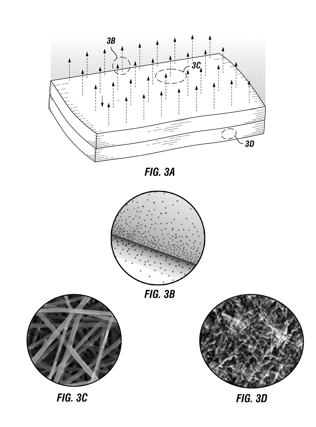
[0175]FIG. 16 through FIG. 34H show various embodiments of electrospun patches useful for the controlled release of growth factors to enhance vascularization.

[0176]Clinical Benefits

[0177]Platforms tailored to release molecules where and when needed;

[0178]Biomimetic material to mimic extracellular matrix mechanical and functional properties to promote and direct tissue formation;

[0179]Guide proliferation and maintenance of stem cells;

[0180]Define an animal model to test a material's properties;

[0181]MSV: burst release; (e.g., the higher the copolymer ratio, the more controlled the release);

[0182]PLGA coating provides a second level of control over the release;

[0183]5% 50:50

[0184]VEGF / PLGA-MSV

[0185]10% 75:15

[0186]PDGF-BB / PLGA-MSV

[0187]By integrating PLGA-MSV in the collagen mats (camouflage) it is possible to ensure their spatial confinement and the preservation of their payload's relea...

example 3

Cardiopatch—Biodegradable, Implantable Patch for Self-directing Autologous Stem Cells to Promote Tissue Regeneration

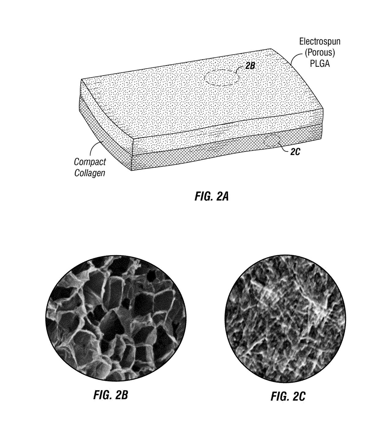
[0196]FIG. 35 through FIG. 43 show various views of exemplary biodegradable, implantable electrospun patches (e.g., a “CardioPatch”) useful for treating conditions of the heart, and in delivering self-directing autologous stem cells to promote tissue regeneration.

[0197]Epidemiology:

[0198]Congenital heart defects (CHD) affect nearly 1% of live births in the United States, equating to nearly 40,000 births per year. Septal wall is the most common and 25% of these will require surgical repair.

[0199]Economic impact:

[0200]In the US alone an estimated $1.8 billion was the net hospital and care costs in 2011. This represented an average of $23,000 expenditures per patient yearly.

[0201]To date, there are no biocompatible cardiac patches (biopatch) capable of growing with the patient, to remodel their structure, and match their function with cardiac tissue.

[0202]“Off the Shelf” ...


PUM

PropertyMeasurementUnit
Fractionaaaaaaaaaa
Fractionaaaaaaaaaa
Lengthaaaaaaaaaa

Abstract

Disclosed are biocompatible, biodegradable, implantable devices for the controlled release of bioactive molecules. In particular embodiments, nanotechnology-based tunable implants are disclosed for 1) localized delivery of analgesics to treat postoperative pain; 2) sustained delivery of growth factors to promote vascularization; and 3) directing tissue regeneration, including the self-direction of autologous stem cells for organ remodeling.

Description

CROSS-REFERENCE TO RELATED APPLICATIONS[0001]The present application claims priority to PCT International Patent Application No. PCT / US2017 / 000017, filed Feb. 22, 2017 (nationalized; Atty. Dkt. No. 37182.192WO01); which claims priority to U.S. Provisional Patent Application No. 62 / 298,407, filed Feb. 22, 2016 (expired; Atty. Dkt. No. 37182.192PV01); the contents of each of which is specifically incorporated herein in its entirety by express reference thereto.STATEMENT REGARDING FEDERALLY SPONSORED RESEARCH OR DEVELOPMENT[0002]Not Applicable.NAMES OF THE PARTIES TO A JOINT RESEARCH AGREEMENT[0003]Not Applicable.BACKGROUND OF THE INVENTIONField of the Invention[0004]The present invention relates generally to the fields of medicine and surgery, and in particular, to implantable drug delivery systems. Disclosed are biocompatible, biodegradable, implantable devices for the controlled release of bioactive molecules. In particular embodiments, nanotechnology-based tunable implants are disc...

Claims


Application Information

Patent Timeline
IPC IPC(8): A61L31/16A61L27/54A61L31/14A61L27/58
CPCA61L31/16A61L27/54A61L31/148A61L27/58A61L17/06A61L2300/604A61L2300/414A61L27/34A61L27/24A61L27/56A61K9/0024A61L2300/402
Inventor TASCIOTTI, ENNIOTARABALLI, FRANCESCAMINARDI, SILVIA
Owner THE METHODIST HOSPITAL
Learn More