Products for repairing cartilage lesions, method of preparation and uses thereof

a cartilage lesions and product technology, applied in the field of cartilage lesions, can solve the problems of limited repair capabilities, increased risk of traumatic lesions of cartilage tissue, and well known cartilage tissu

Inactive Publication Date: 2017-04-27
STEMMATTERS BIOTECHA E MEDICINA REGENERATIVA
View PDF0 Cites 2 Cited by
  • Summary
  • Abstract
  • Description
  • Claims
  • Application Information

AI Technical Summary

Problems solved by technology

Cartilage tissue is well known by those skilled in the art to have very limited repair capabilities when injured.
The concomitant increase in life expectancy worldwide and physical activity throughout life time, in which sport activities play an important role as they are practiced up to seniority, increases risk for traumatic lesions of the cartilage tissue, as well as increases likelihood of cartilage wear by use.
As a consequence, cartilage lesions can lead to physical impairment and interruption of normal daily activities, causing absence from work and/or school and premature abandonment of sports activity.
First line of treatment involves long-term physical therapy and drugs, which render limited efficacy.
Currently, there is yet no fully clinically acceptable therapeutic option for focal cartilage lesions.
Furthermore, tissue availability for transplant constitutes a major limitation, especially in large cartilage defects.
However, ACI application may be inadequate in certain scenarios because of anatomic factors, and difficulty of fixation, in degenerative defects, of the periosteal flap or collagen sheets to retain the chondrocyte suspension.
Other potential complications include periosteal hypertrophy, ablation, uneven cell distribution, and loss of cells into the joint cavity resulting in the need of repetition of surgery in up to one third of the patients.
However, even this therapy presents several performance drawbacks resulting in surgical complications, which normally leads to repetition of surgery in 25 to 36% of the ACT treated patients [Harris, J. D., et al., Osteoarthr Cartilage, 2011.
However, it is noted that ionic-crosslinked hydrogels made from methacrylated gellan gum with high substitution degree have poor mechanical properties.
This aspect represents a limitation in terms of the need for additional equipment and time required for obtaining homogeneous solutions of the materials.
Furthermore, this temperature is incompatible for applications in animals and humans, wherein physiological temperature is approximately 37° C. Ideally, the methacrylated gellan gum should be readily soluble in water between room temperature and 37° C.
While photo-crosslinking prom

Method used


Image

  • Products for repairing cartilage lesions, method of preparation and uses thereof
  • Products for repairing cartilage lesions, method of preparation and uses thereof
  • Products for repairing cartilage lesions, method of preparation and uses thereof

Examples


example 1

on of Tissue Engineering Product Composition for Cartilage Repair

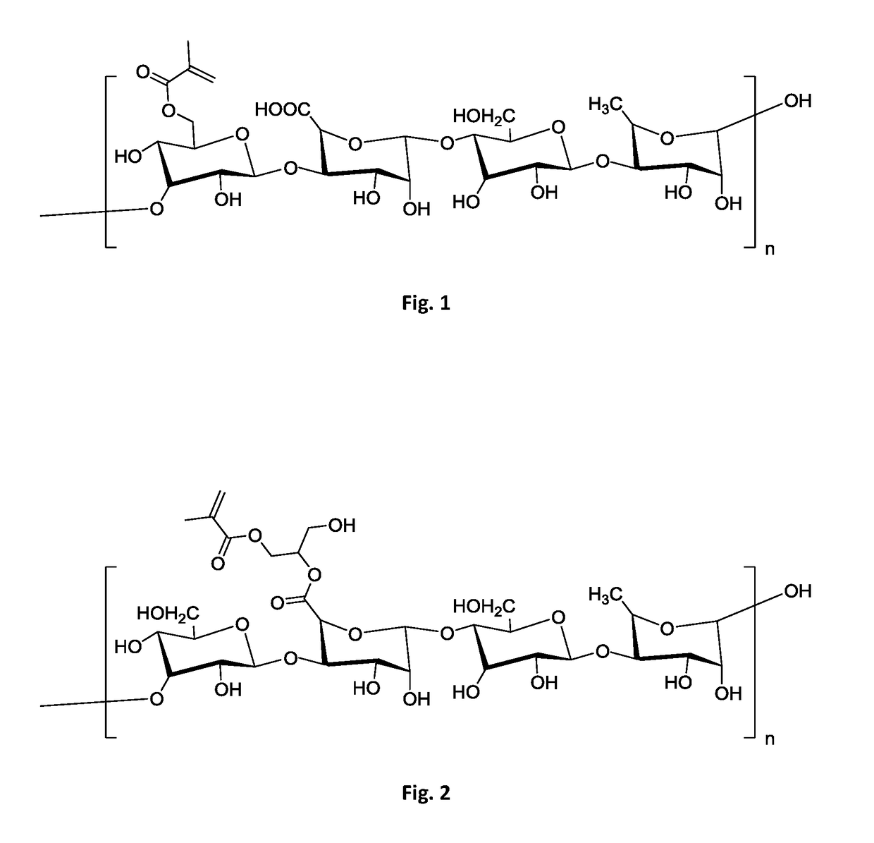
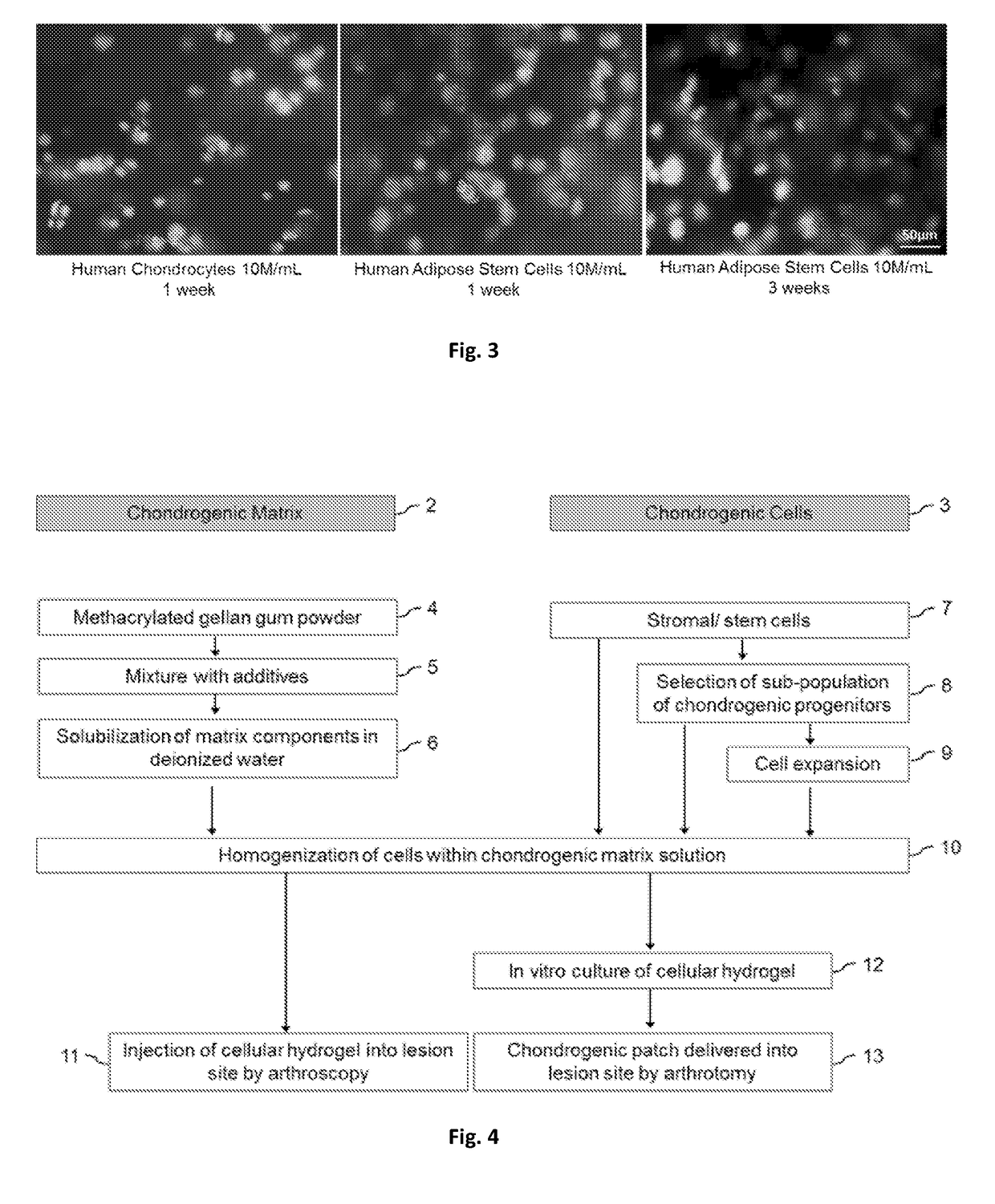
[0104]An aseptic environment was set to prepare the chondrogenic composition. Chondrogenic matrix was prepared by sourcing 20 mg of methacrylated gellan gum powder with a methacrylation degree between 1.5 and 5%. Quality control ensured absence of any microbial contamination, as well as ensuring levels of mycoplasma and endotoxins below limits acceptable for therapeutic use. An aqueous solution was prepared by homogenizing said powder with sterile deionized water, yielding a 2% w / V solution. Homogenization was performed at 37° C. with mild agitation. Chondrogenic cells were prepared by sourcing 10 million human stromal / stem cells, at passage 1-2, from a master cell bank. Said human stromal / stem cells were isolated in xeno-free conditions from adipose tissue of a qualified donor. The donor sample was qualified as for absence of bloodborne pathogens and absence of known medical conditions. Cells were qualified for presen...

example 2

Development of Hyaline Cartilage by the Use of Disclosed Composition

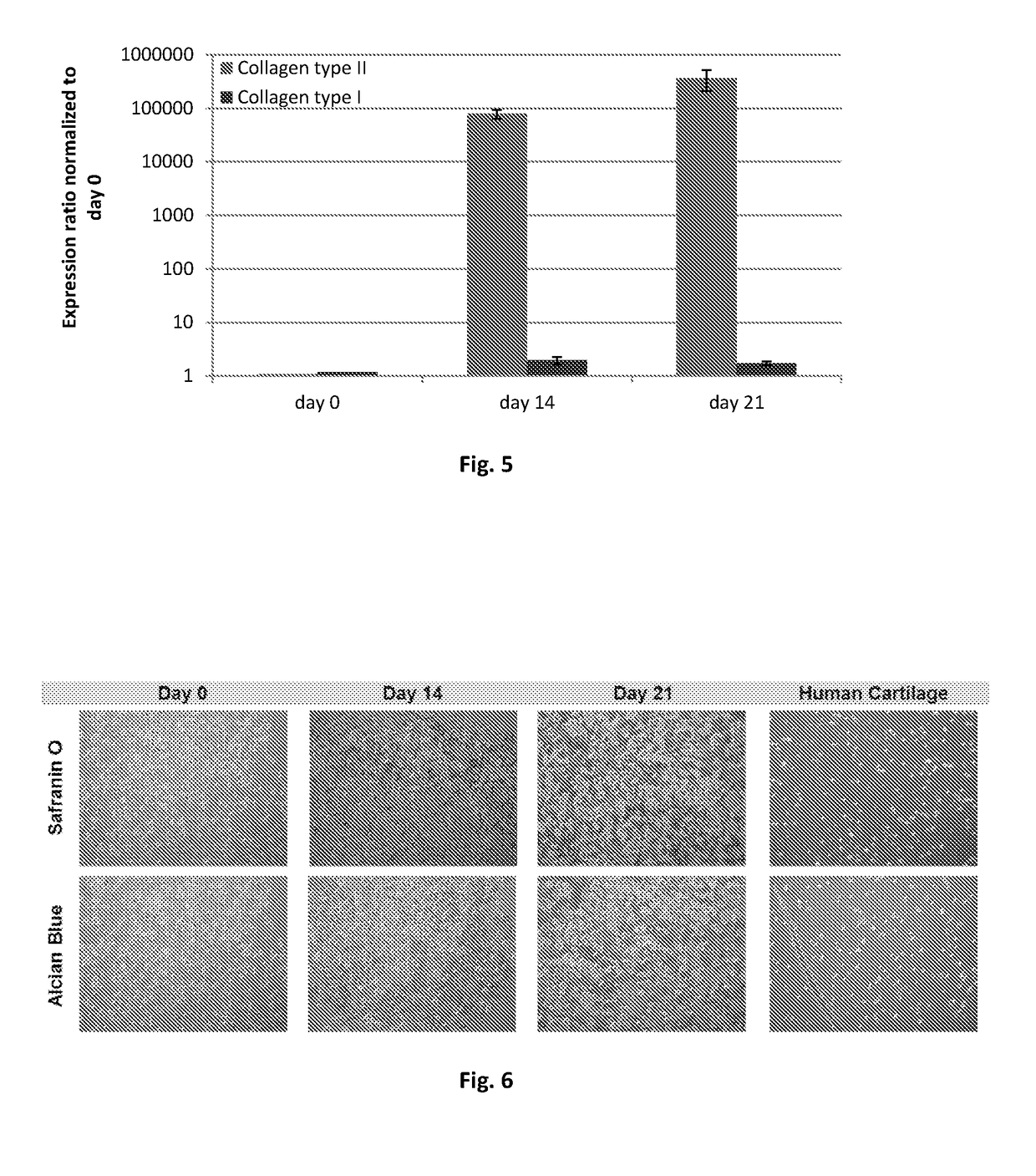
[0105]Healthy hyaline articular cartilage is evaluated by the composition of its extracellular matrix, which includes mainly collagen type II and glycosaminoglycans. When fibrous cartilage is formed, the composition of extracellular matrix shifts, giving rise to molecules such as collagen type I that render less elasticity to the tissue, thereby becoming less capable to withstand mechanical demands of the joint. This procedure may be applied to the evaluation of any composition of this invention.

Materials and Methods

[0106]The cartilage repair composition, as described in example 1, was cultured in vitro for 21 days, exposed to chondrogenic growth factors. In vitro-developed grafts were collected for histological assessment according to standard procedures. Safranin O and alcian blue stainings were performed to detected cartilage glycosaminoglycans. Other grafts were used for quantitative determination collagen type ...

example 3

epair of Rabbit Hyaline Cartilage Lesion by the Use of Disclosed Composition

[0111]The performance and efficacy of disclosed composition and method for treatment of focal cartilage lesions was assessed in a rabbit model.

Materials and Methods

[0112]A rabbit model was used to test the efficacy of the disclosed composition and method on the repair of cartilage lesions. A focal articular cartilage lesion was induced to the animal's knee by the use of a biopsy punch and curette. Lesions were immediately treated either with the preferred embodiment described in example 1, or adopting a current standard of care surgical method—microfracture. As control, lesions were left untreated. An 8 week repair period was allowed, after which articular cartilage samples were harvested for histological analysis. Safranin O / fast green staining was performed to identify status of lesion repair.

Results

[0113]FIG. 7 represents microscopic images of rabbit articular cartilage sections stained with safranin O / fa...


PUM

PropertyMeasurementUnit
Fractionaaaaaaaaaa
Fractionaaaaaaaaaa
Fractionaaaaaaaaaa

Abstract

Products for repairing cartilage lesions, method of preparation and uses thereof The present disclosure provides products and methods of preparation thereof, said products comprising a matrix of methacrylated gellan gum having a methacrylation degree between 1.5 and 6%, cartilage forming cells and a physiologically acceptable ionic solution containing cations, for application in tissue engineering and regenerative medicine, in particular cartilage lesions.

Description

TECHNICAL FIELD[0001]The present disclosure relates to products and preparation methods for the treatment of tissues, in particular cartilage lesions, by means of tissue engineering and regenerative medicine. A composition includes a matrix and cartilage forming cells.BACKGROUND ART[0002]Articular cartilage tissue is composed by one single cell type—chondrocytes, a dense extracellular matrix which constitutes 20% of the tissue, while the remaining composition of cartilage (approximately 80%) is water. It completely lacks nervous and vascular systems, which are those mostly involved in tissue repair mechanisms. Cartilage tissue is well known by those skilled in the art to have very limited repair capabilities when injured.[0003]The concomitant increase in life expectancy worldwide and physical activity throughout life time, in which sport activities play an important role as they are practiced up to seniority, increases risk for traumatic lesions of the cartilage tissue, as well as i...

Claims


Application Information

Patent Timeline
IPC IPC(8): A61L27/20A61L27/26A61L27/38C08B37/00A61L27/52
CPCA61L27/20C08B37/006A61L27/52A61L2400/06A61L27/26A61L2430/06A61L27/3852A61L27/3834
Inventor SOUSA, RUI PEDRO ROMERO AMANDI DECORREIA, CRISTINAVILELA GOMES, CARLOS ALBERTODA SILVA MORAIS, ALAIN JOSEFREIRE GERTRUDES, ANA CATARINAXAVIER CARLOS DOS SANTOS, TIRCIA SUSETEANTUNES CORREIA DE OLIVEIRA, JOAQUIM MIGUELDUARTE COELHO DO SAMEIRO ESPREGUEIRA MENDES, JOAO DUARTEGON ALVES DOS REIS, RUI LUIS
Owner STEMMATTERS BIOTECHA E MEDICINA REGENERATIVA
Why Eureka
Try Eureka