
Diagnostic method for colorectal cancer

Inactive Publication Date: 2015-10-08
ORIGIN SCI
View PDF1 Cites 7 Cited by
  • Summary
  • Abstract
  • Description
  • Claims
  • Application Information

AI Technical Summary

Benefits of technology

The patent provides methods for diagnosing and monitoring the progress of colorectal cancer in patients using a sample of the colorectal mucosa obtained from the patient. The methods involve measuring the levels of certain proteins in the sample and comparing them to levels in control samples or samples from patients with colorectal cancer. A difference in protein levels can indicate a diagnosis of colorectal cancer, while a similarity in protein levels can suggest a diagnosis. The methods can be used to monitor the effectiveness of therapy for colorectal cancer. The patent also includes a kit for detecting and quantifying the proteins of the colorectal mucosa.

Problems solved by technology

It can be difficult to diagnose, particularly because symptoms are often left unnoticed until a tumor is well advanced.
Fecal occult blood testing can give a general indication of bleeding in the bowel, but it is not able to provide an early diagnosis of colorectal cancer or indicate the presence of polyps which may develop into colorectal cancer.
Sigmoidoscopy and colonoscopy techniques are usually used to screen for colorectal cancer, but these methods can be time consuming and expensive.
Individuals with Inflammatory Bowel Disease, including Ulcerative Colitis and Crohn's Disease, have an increased risk of developing colorectal cancer.

Method used


Image

  • Diagnostic method for colorectal cancer
  • Diagnostic method for colorectal cancer
  • Diagnostic method for colorectal cancer

Examples


example 1

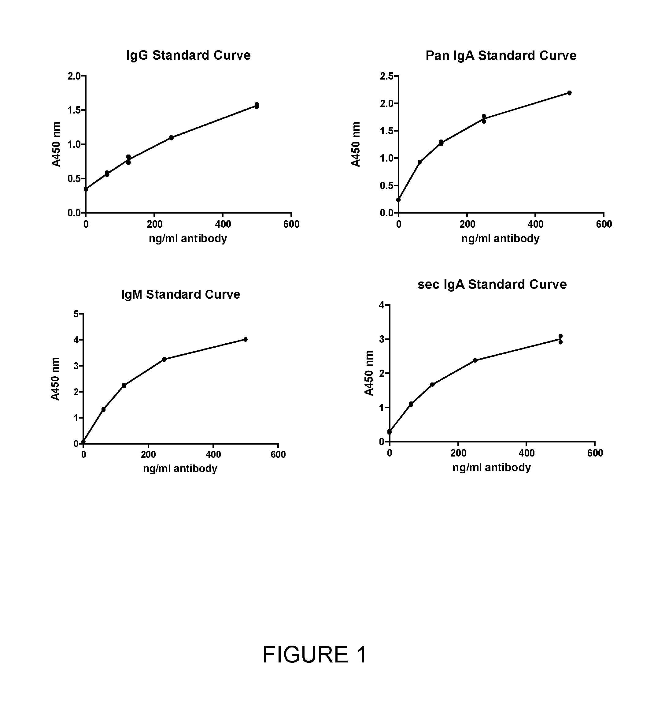
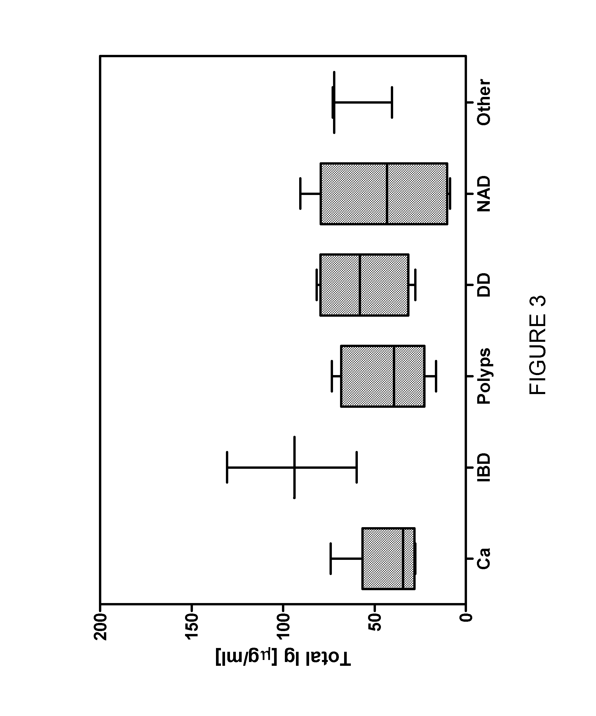
Detecting Total Concentration of Antibodies in Clinical Samples Using an Anti-Human Antibody ELISA

[0113]Materials and Methods

ReagentsStockComponentSupplierconcentrationCarbonate-bicarbonate buffer tabletsSigma1 tab = 100 mlPhosphate buffered saline (PBS)Fisher Scientific10x Tween 20Fisher Scientific1xBSASigma1xMAb anti-human IgM FcStratech (HRL)2 mg / mlMAb anti-human IgA Fc PANStratech (HRL)2 mg / mlMAb anti-human IgG FcStratech (HRL)2 mg / mlMAb anti-human SecIgA - biotinStratech (HRL)1 mg / mlMAb anti-human IgA Fab PAN -Stratech (HRL)1 mg / mlbiotinMAb anti-human IgG Fd - biotinStratech (HRL)1 mg / mlMAb anti-human IgM Fc - biotinStratech (HRL)1 mg / mlIgA from human colostrumsMP Biomedicals5 mg / mlIgM from human serumSigma0.8 mg / ml  IgG from human serumSigma6 mg / mlExtrAvidin ™-PeroxidaseSigma1.23 mg / ml  Enhanced K-Blue TMB SubstrateSkybio1xHClFisher Scientific36% (11.65M)

BuffersBufferCompositionCoat buffer50 mM Na2CO3, pH 9.6PBS11.9 mM Na2HPO4, 11.9 mM KH2PO4, 137 mMNaCl, 2.7 mM KClWash buffer...

example 2

Anti-Carbohydrate ELISA with IgA Detection

[0148]Materials and methods

[0149]Reagents

ComponentSupplierStock concentrationCarbonate-bicarbonateSigma1tab = 100 mlbuffer tabletsLNFP II-BSA / Lewisa-Dextra Laboratories1mg / ml in DPBSBSA (lactose spacer)LNFP III-BSA / LewisX-Dextra Laboratories1mg / ml in DPBSBSA (lactose spacer)LNFP I-BSA / BloodDextra Laboratories1mg / ml in DPBSgroup H TIT-antigen-HSADextra Laboratories1mg / ml in DPBSTn-antigen-HSADextra Laboratories1mg / ml in DPBSLewisx-BSADextra Laboratories1mg / ml in DPBS3′-Sialyl Lewisx-BSADextra Laboratories1mg / ml in DPBSBlood group A-BSADextra Laboratories1mg / ml in DPBSBlood group B-BSADextra Laboratories1mg / ml in DPBSGalα1-3Galβ1-Dextra Laboratories1mg / ml in DPBS4GlcNAc-BSABSA (coat)Sigma-AldrichN / AHSA (coat)Sigma-AldrichN / ABSA (block)Sigma-AldrichN / ATween 20Fisher Scientific1xPhosphate buffered salineFisher Scientific10x (PBS)MAb anti-humanStratech (HRL)1mg / mlSecIgA - biotinExtrAvidin ™-Sigma1.23mg / mlPeroxidaseEnhanced K-Blue TMBSkybio1xSubs...

example 3

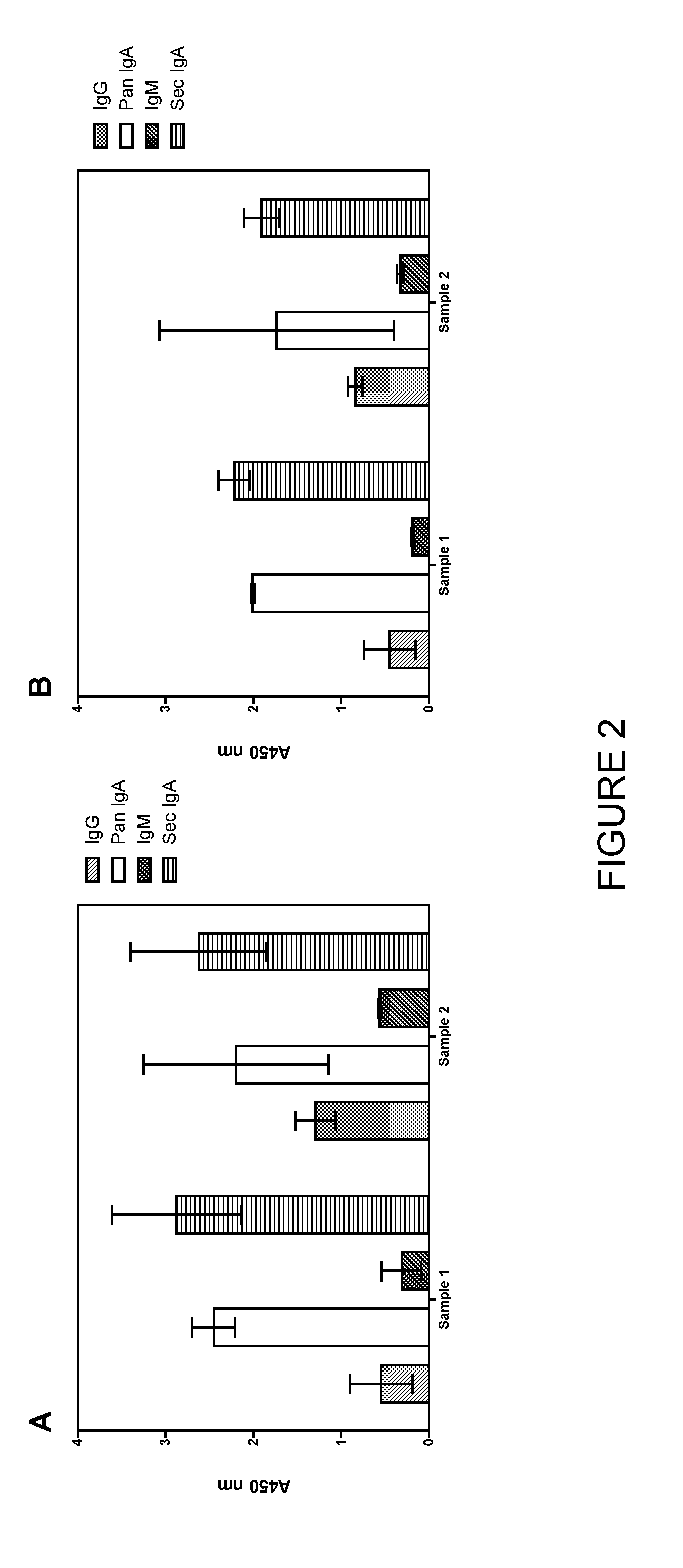
Anti-Carbohydrate ELISA with IgA (Pan and Secretory), IgM and IgG with Samples from Healthy Volunteers

[0173]Materials and Methods

[0174]Reagents

[0175]Same as described in Example 2, above, with the addition of the following detection antibodies:

ComponentSupplierStock concentrationMab anti-human PANIgAStratech (HRL)1 mg / mlMab anti-human IgMStratech (HRL)1 mg / mlMab anti-human IgGStratech (HRL)1 mg / ml

[0176]Buffers

[0177]Same as described in Example 1, above.

[0178]Samples

[0179]The colorectal mucosal samples were taken from two healthy volunteers (volunteer (a) and volunteer (b)). A second sample was taken from volunteer (b) 14 weeks after the first sample and the results compared.

[0180]Protocol

[0181]Same as described in Example 2, above.

[0182]Results

[0183]A sample was collected from two healthy volunteers and tested for anti-IgA, anti-IgM and anti-IgG antibodies to 10 carbohydrate antigens (T-antigen, Tn-antigen, Lewis X, α-gal, Lewis A (lactose spacer), sialyl Lewis X, Lewis X (lactose s...


PUM

PropertyMeasurementUnit
Concentrationaaaaaaaaaa
Levelaaaaaaaaaa

Abstract

The present invention relates to a method of diagnosing patients with colorectal cancer by quantifying the level of one or more proteins, for example immunoglobulins such as IgM, IgG, IgA (total and / or secretory IgA), in a sample of the colorectal mucosa. The invention also relates to methods of monitoring efficacy of colorectal cancer therapy and to kits for diagnosing colorectal cancer by using the method described herein.

Description

FIELD OF THE INVENTION[0001]The present invention relates to a method of diagnosing patients with colorectal cancer by quantifying the level of one or more proteins in a sample of the colorectal mucosa. The invention also relates to methods of monitoring efficacy of colorectal cancer therapy and to kits for diagnosing colorectal cancer by using the method described herein.BACKGROUND OF THE INVENTION[0002]Colorectal cancer is thought to be the third most common cancer in the world, with almost 60% of cases being diagnosed in the developed world. It can be difficult to diagnose, particularly because symptoms are often left unnoticed until a tumor is well advanced. However, early diagnosis of colorectal cancer is important in order to improve a patient's chances of survival.[0003]Typical methods of diagnosis of colorectal cancer include fecal occult blood testing, sigmoidoscopy and colonoscopy. Fecal occult blood testing can give a general indication of bleeding in the bowel, but it is...

Claims


Application Information

Patent Timeline
IPC IPC(8): G01N33/574
CPCG01N33/57419G01N33/6854G01N2400/00G01N2800/52
Inventor BOOTH, JODIE LOUISE
Owner ORIGIN SCI
Learn More