
Method and system for assessing preterm birth and other pathologies

a preterm birth and other pathology technology, applied in the field of medical imaging, can solve the problems of premature birth, serious morbidity in surviving infants, and remains elusiv

Inactive Publication Date: 2015-03-19
THE JOHN HOPKINS UNIV SCHOOL OF MEDICINE +1
View PDF0 Cites 5 Cited by
  • Summary
  • Abstract
  • Description
  • Claims
  • Application Information

AI Technical Summary

Benefits of technology

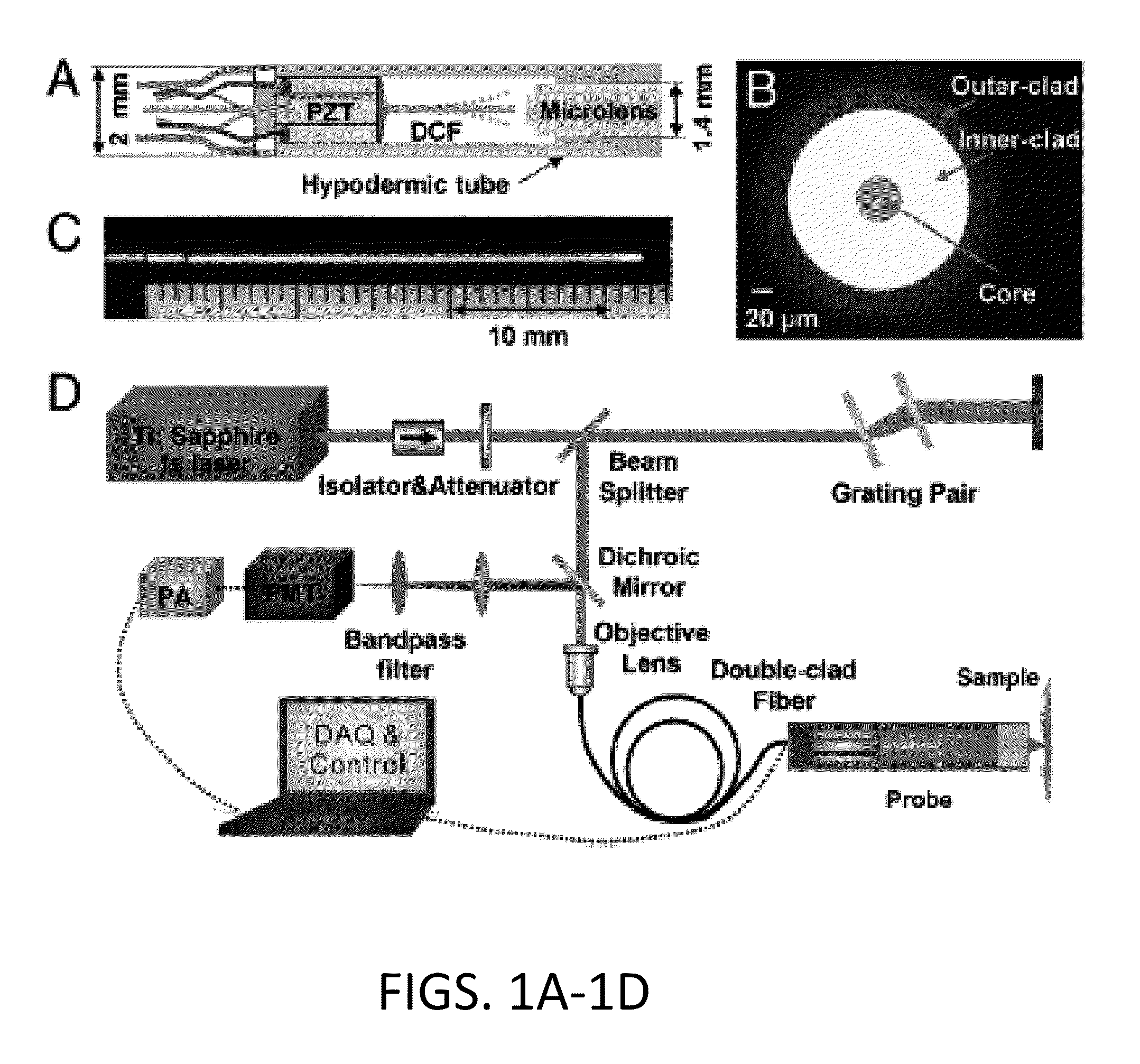
The present invention provides a method for assessing pre-term birth in real-time using a fiber-optic scanning second harmonic generation (SHG) endoscopy imaging system. The method involves obtaining SHG endoscopy images of cervical collagen and determining collagen fiber morphologies, such as diameter, volume, porosity, and bending angle, to determine a risk of pre-term birth. The SHG endoscopy system uses a short pulsed laser source, fiber optic scanning endoscope, light detection unit, and computer control. The technical effect is the ability to accurately assess pre-term birth risk in real-time, which can improve pre-natal care and outcomes for newborns.

Problems solved by technology

Abnormal or inappropriately timed cervical remodeling can lead to premature birth.
Preterm birth (PTB), which accounts for 12.7% of all births in the United States, is the second leading cause of infant mortality and often leads to serious morbidity in surviving infants.
Despite considerable research, the cause of PTB in 50% of cases remains elusive, and diagnostic methods to detect women at risk of PTB are limited.
Translation of cervical SHG microscopy for clinical use in vivo represents a formidable engineering challenge, which requires miniaturization of a bench-top microscope down to a small imaging probe applicable for minimally invasive imaging of the cervix.
Several critical technological challenges and engineering building blocks for developing a high-performance SHG endomicroscopy technology exist.
In addition to the challenges in effective excitation pulse delivery, SHG signal collection and miniature beam scanner, one significant challenge for an SHG imaging endomicroscope is the more restrictive demand for low chromatic aberration in the micro-objective lens, considering the large wavelength separation between the excitation and emission in SHG imaging (which is typically much larger than the separation in two-photon fluorescence imaging).

Method used


Image

  • Method and system for assessing preterm birth and other pathologies
  • Method and system for assessing preterm birth and other pathologies
  • Method and system for assessing preterm birth and other pathologies

Examples


Embodiment Construction

[0022]The presently disclosed subject matter now will be described more fully hereinafter with reference to the accompanying Drawings, in which some, but not all embodiments of the inventions are shown. Like numbers refer to like elements throughout. The presently disclosed subject matter may be embodied in many different forms and should not be construed as limited to the embodiments set forth herein; rather, these embodiments are provided so that this disclosure will satisfy applicable legal requirements. Indeed, many modifications and other embodiments of the presently disclosed subject matter set forth herein will come to mind to one skilled in the art to which the presently disclosed subject matter pertains having the benefit of the teachings presented in the foregoing descriptions and the associated Drawings. Therefore, it is to be understood that the presently disclosed subject matter is not to be limited to the specific embodiments disclosed and that modifications and other ...


PUM


Abstract

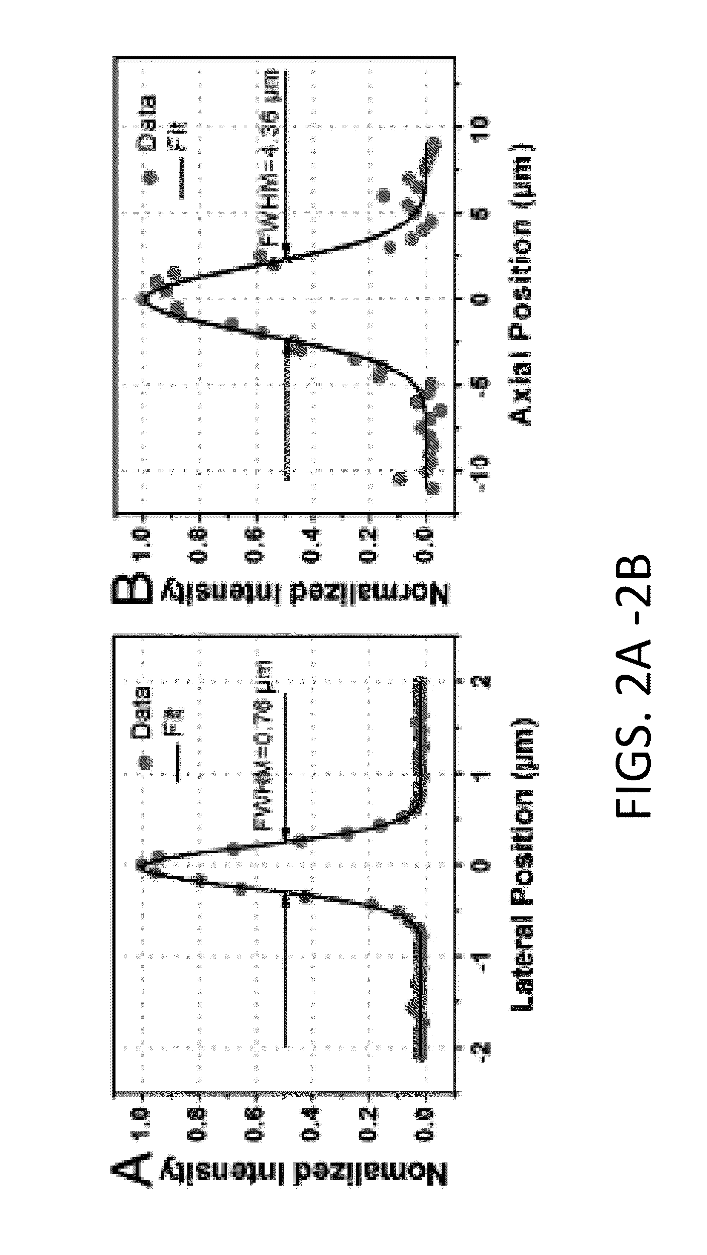
The present invention is directed to an all-fiber-optic scanning endomicroscope capable of high-resolution second harmonic generation (SHG) imaging of biological tissues. The endomicroscope has an overall 2.0 mm diameter and consists of a single customized double-clad fiber, a compact rapid two-dimensional beam scanner, and a miniature compound objective lens for excitation beam delivery, scanning, focusing, and efficient SHG signal collection. Endomicroscopic SHG images of murine cervical tissue sections at different stages of normal pregnancy reveal progressive, quantifiable changes in cervical collagen morphology with resolution similar to that of bench-top SHG microscopy. A device according to an embodiment of the present invention can also be used to assess other pathologies, such as cancer. fibrosis, and inflammation. The present invention allows for diagnosis, monitoring of the effect of therapeutics, and surgical or interventional guidance. The present invention enables visualization of histology in-vivo, in the patient, and is label-free.

Description

CROSS REFERENCE TO RELATED APPLICATIONS[0001]This application claims the benefit of U.S. Provisional Patent Application No. 61 / 863,022 filed on Aug. 7, 2013, which is incorporated by reference, herein, in its entirety.GOVERNMENT SUPPORT[0002]This invention was made with government support under grant no. CA153023 awarded by the National Institutes of Health. The government has certain rights in the invention.FIELD OF THE INVENTION[0003]The present invention relates generally to medical imaging. More particularly, the present invention relates to a method and system for assessing preterm birth and other pathologies using medical imaging.BACKGROUND OF THE INVENTION[0004]The cervix is a connective tissue-rich structure just caudal to the uterus in female mammals. Appropriate remodeling of the cervix during gestation is an essential component of the birth process. The cervix must remain closed during pregnancy to maintain the fetus in the womb, and then open sufficiently to allow passag...

Claims


Application Information

Patent Timeline
IPC IPC(8): A61B5/00A61B1/06A61B5/107A61B1/00A61B1/07A61B1/303
CPCA61B5/435A61B1/07A61B1/303A61B5/0084A61B5/1076A61B5/7282A61B5/7275A61B1/00009A61B1/0669A61B5/0062A61B5/1079A61B5/4331A61B1/00172A61B1/043A61B1/063
Inventor LI, XINGDEZHANG, YUYINGLIANG, WENXUANMAHENDROO, MALALUBY-PHELPS, KATEAKINS, MEREDITH
Owner THE JOHN HOPKINS UNIV SCHOOL OF MEDICINE
Learn More