On the other hand, IHC and IHF cannot be immediately applied to sample tissues but some pre-
processing stages are needed.
One of the immediate problems is the long duration of the
processing cycle.
This is currently a big obstacle of current IHC processes, since the needed time period does not allow analysis to be done during
surgical interventions.
Unfortunately, until now, there has not been a technique presented to verify that the tumour is completely clean.
When counted, the number of surgeries varies from 1 to 3, which increases risks, costs and
anxiety for the patients, as well as a significant loss of health resources like doctors' time and
surgery room availability.
First, in the cryo-fixation procedure,
tissue preservation and archiving are more costly due to the needed equipment.
Apart from the intra-operative aspect, the accuracy of the obtained results with any technology until now is limited, when dealing with cases requiring quantitative biomarker
expression analysis using the extent of the obtained
signal by
immunohistochemistry, as required during certain assays.
Conventional techniques can produce ambiguous results up to 20% cases when such semi-quantitative analysis is required, and a final diagnostic result cannot be achieved using
immunohistochemistry alone.
Therefore, current standard is to subject these cases to a subsequent
genetic analysis (
in situ hybridization) in order to achieve a final diagnostic outcome, adding substantial cost and time (a few days) to the diagnostic process.
The inaccuracy of the quantitative immunohistochemical analysis has its origins in the intensity of an immunohistochemical
signal, which is not necessarily proportional to the extent of
antigen expression due to non-specific binding reactions, as well as unpredictable effects of
tissue degeneration, variations in tissue fixation,
paraffin embedding, and heat-induced
epitope retrieval.
This originates from long
diffusion times, lack in precision of controlling and dosing of reagents, as well as limited fluidic exchange rates.
In addition, long
assay and
antibody exposure times may result in significant adsorption and non-specific binding of the antibodies, so that the
resultant immunohistochemical
signal is no longer a linear function of the target biomarker concentration on the tissue.
This problem exists in all settings where an immobilized target is present on the surface, and one or more
detector reagent binds to this target at a rate limited by the
diffusion speed of the
detector reagents.
Plus, the steric hindrance mechanisms can also contribute to this non-proportionality and compromises an eventual
quantitative assay.
State-of-the-art automated equipments have a few other drawbacks in addition to the intrinsic problems of long
process duration and limited accuracy.
Modern commercial automated IHC are bulky, supplied either in a bench or placed at the bench top.
Therefore, they are far from being portable and hand-held.
In general clinics with a low budget and those that are located at remote places do not have the necessary infrastructure, equipment and expertise to be able to perform such kind of diagnosis.
In fact, it is extremely expensive to form and maintain such a laboratory for a small sized clinic, requiring around 1M CHF investment on infrastructure and equipment, and more than 300K CHF per year for trained personnel.
One additional major obstacle caused by the current structure of a laboratory dictated by state-of-the-art equipment is the customization problem, which appears in particular when using for new biomarker discoveries and related research.
In addition, the central facilities
resist such customization because either they are overloaded with the current diagnosis work or such customization may affect the later reproducibility.
However, when thought the large number of trials required to validate results and requirement for the reproducibility, the total time needed for manually completing such studies can span a few years, significantly affecting the total research and development costs of
biomarker discovery.
However, in none of these studies there had been an implication that a microfluidic approach results in an increase in accuracy of quantitative analysis and a decrease ambiguous diagnostic results obtained by such analyses.
Unfortunately, this device showed a limited analysis speed and detection area.
The
system was unable to hold high pressures, resulting in a maximum operational volumetric flow rate around 50 nL / s.
The cumbersome
assembly and disassembly of the
system (manual integration) significantly increased analysis time and
dead volume.
Also, only part of the tissue slice could be exposed (less than a few 10% of the surface), thereby limiting the TS detection area.
On the other hand, this device suffered from a number of problems that largely compromise the accuracy of quantitative analysis and its low-cost commercialization.
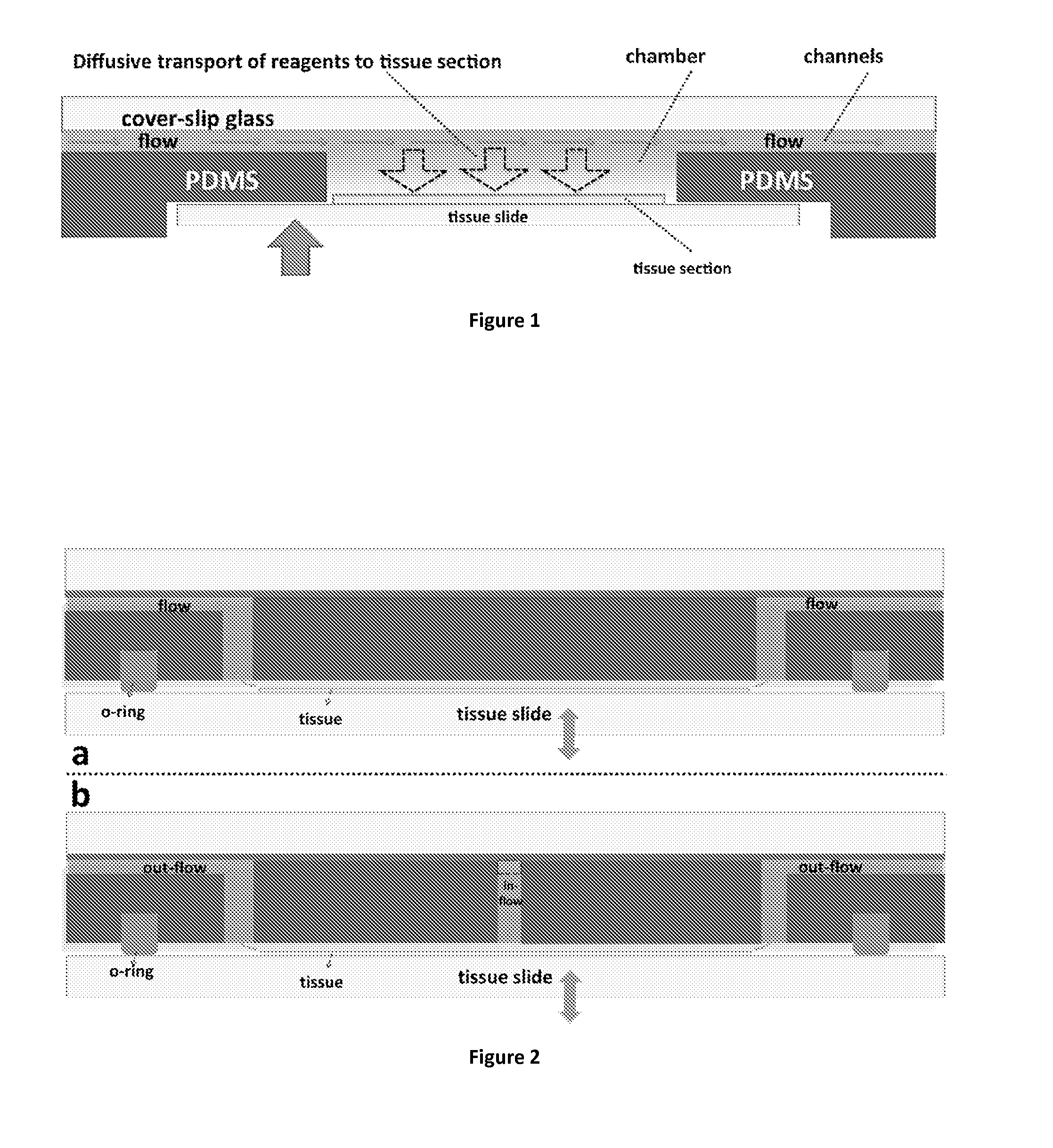
In addition, the design dictates that the chamber height should always be higher than the height of the microfluidic channels, and this condition renders the limiting role of diffusion in the transport of the antibodies even more significant.
More strikingly, the slow diffusion-limited transport of the protocol reagents to the
tissue surface compromises the proportionality between the obtained signal and the extent of
antigen expression on the
tissue surface, which makes a successful
quantitative assessment of the immunohistochemical signal impossible.
Using standard
fluorescent imaging equipment and commercial fluorophores, it would not be possible to detect this signal.
Nevertheless, both the fluorophores and
microscopy equipment for time-resolved markers (lanthanides) are very expensive and non-standard, and conjugated diagnostic antibodies are commercially not available.
As a consequence, the requirement for time-resolved
microscopy constitutes another obstacle preventing successful and low-cost commercial implementation of this device, which, therefore cannot operate when employing standard
staining reagents like fluorophores and chromogens.
Apart from the design-related problems that are listed above (paragraphs 0016-0017 and cited document), there are also a number of drawbacks due to the use of PDMS as a
structural material.
The relatively low Young's modulus of PDMS makes the channels susceptible to substantial deformation under higher fluidic pressures.
Again, due to the low Young's modulus of PDMS, the force required for better sealing of the tissue slide can easily deform these channels, up to an extent that they are blocked and the operation of the device becomes impossible.
These variations in the design dimensions and flow-rates due to easy deformation of the channels can introduce variations in the
resultant IHC signal, and, when used for diagnosis, can largely compromise reproducibility of the results.
In addition, the thermal properties of PDMS prevent the use of temperatures above 70° C., and PDMS is not chemically compatible with the many reagents, line
Xylene, that may be involved in IHC protocols.
Another drawback of this device is that it only accepts non-standard
tissue section slide shapes, which constitutes a customization and hence a cost problem in commercialization issue of this structure as a diagnostic device.
To conclude, the described
system in paragraphs 0016-0018 only improves the protocol time of the immunohistochemical
assay, but this at the cost of using time-resolved
fluorescence, which is an expensive and mostly inaccessible method in terms of required materials and infrastructure.
Moreover, the
assay time minimization does not eliminate diffusion-controlled transport of reagents to the
tissue surface, and the ability to perform quantitative analysis that can be done with this system does not differ from a conventional setting: no quantification of tissue biomarkers is possible.
Last but not least, the low Young's modulus of the system highly compromises the reproducibility due to possible deformations in the microfluidic structures that form the system.
Moreover, the device can
stain about 1.5% of the TS area, which is a drawback for generalization of the technique.
Although authors have shown, for the specific case of
breast cancer, that even for this small area there is about 85% correlation with a totally stained TS, a sufficient correlation cannot be achieved.
It is also a cumbersome work to realize this correlation study for each different case.
In addition, the lab-on-a-
chip IHC processors either semi-manual as in the case of MMIHC where only primary
antibody incubation is done on-
chip, can
stain only a proportion of the TS or has high
reagent costs.


