
System and Method for Magnetic Resonance Imaging Based Respiratory Motion Correction for PET/MRI

a technology of magnetic resonance imaging and respiratory motion correction, applied in image enhancement, instruments, applications, etc., can solve the problems of low temporal resolution, difficult application of breath holding techniques, and often degrading image data of pets

Active Publication Date: 2014-12-04
SIEMENS HEALTHCARE GMBH
View PDF9 Cites 32 Cited by
  • Summary
  • Abstract
  • Description
  • Claims
  • Application Information

AI Technical Summary

Benefits of technology

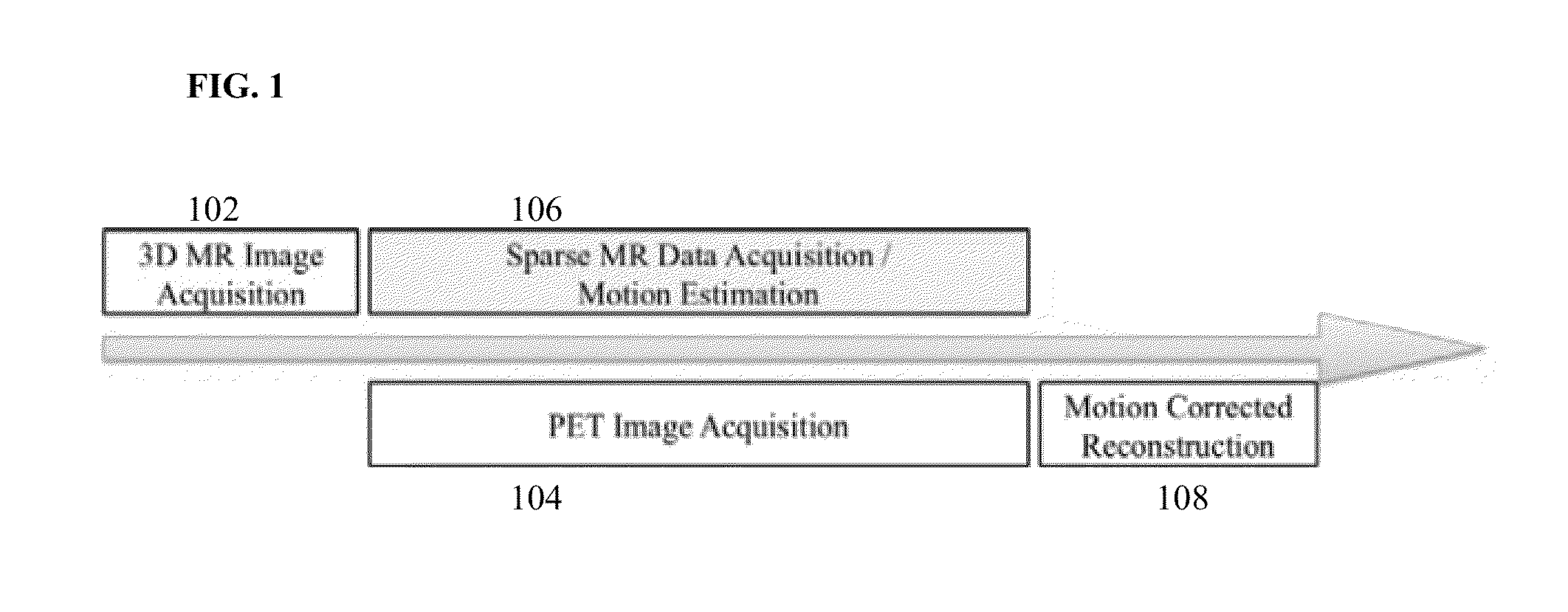
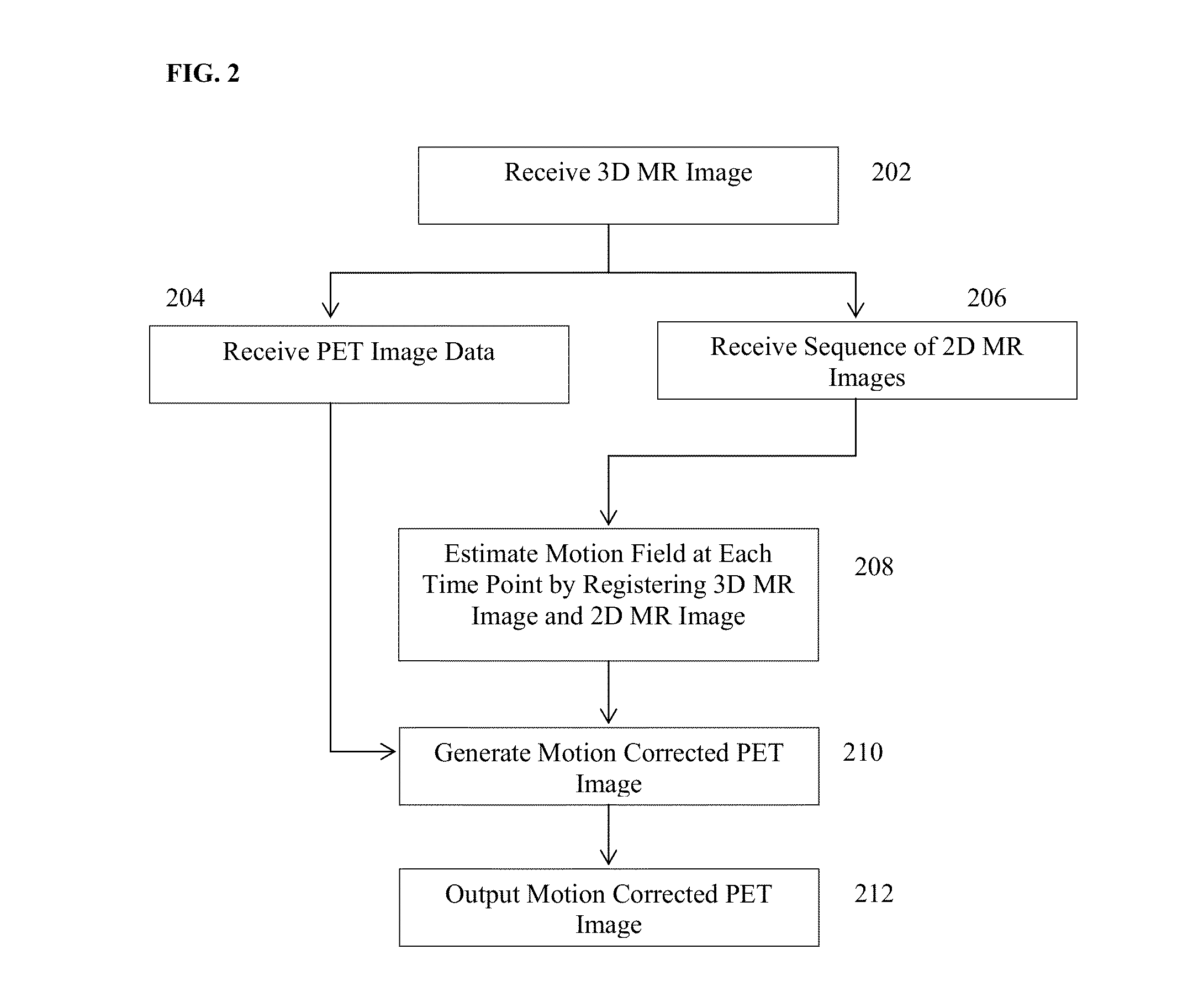
The present invention provides a method and system for MRI based motion correction in a PET image. This can be used for motion correction of a PET image acquired in a PET / MRI image acquisition system. The invention involves acquiring a series of dynamic 2D MR images during the PET image data acquisition, and registering these images against a static 3D MRI to estimate a motion field. This motion field can then be used in PET image reconstruction for motion correction of the PET image data. The technical effect of this invention is to improve the accuracy and reliability of PET image data, which can lead to better outcomes for patients.

Problems solved by technology

However, PET image data often suffers from image degradation due to patient motion during the PET image data acquisition.
Due to the long acquisition time of PET (typically one to three minutes per bed position), breath holding techniques are difficult to apply.
However, such respiratory gating techniques assume that respiratory motions in the same breathing phase are identical across different breathing cycles, which is not true due to the fact that breathing motion patterns can be irregular, especially when the patient is under stress, anxiety, and / or pain.
However, 4D MR image data with a high spatial resolution typically has a very low temporal resolution, which limits its usefulness for motion estimation.
However, due to the tradeoff between spatial and temporal resolution of MR imaging, all of the proposed methods rely on gating for 3D MRI acquisition.
Accordingly, these methods suffer from the same drawbacks as other respiratory gating techniques.

Method used


Image

  • System and Method for Magnetic Resonance Imaging Based Respiratory Motion Correction for PET/MRI
  • System and Method for Magnetic Resonance Imaging Based Respiratory Motion Correction for PET/MRI
  • System and Method for Magnetic Resonance Imaging Based Respiratory Motion Correction for PET/MRI

Examples


Embodiment Construction

[0012]The present invention provides a method and system for MRI based motion correction in a PET image. Embodiments of the present invention are described herein to give a visual understanding of the motion correction method. A digital image is often composed of digital representations of one or more objects (or shapes). The digital representation of an object is often described herein in terms of identifying and manipulating the objects. Such manipulations are virtual manipulations accomplished in the memory or other circuitry / hardware of a computer system. Accordingly, is to be understood that embodiments of the present invention may be performed within a computer system using data stored within the computer system.

[0013]Embodiments of the present invention provide a method of PET motion correction that utilizes a series of 2D MR images with a respiratory motion model for respiratory motion estimation. The use of 2D MR images for motion estimation instead of 3D MR images is advan...


PUM


Abstract

A method and system for magnetic resonance imaging (MRI) based motion correction in position emission tomography (PET) images is disclosed. A static 3D magnetic resonance (MR) image of a patient is received. PET image data of the patient and a series of 2D MR images of the patient acquired simultaneous to the acquisition of the PET image data are received. A 3D+t motion field is estimated by registering the series of 2D MR images acquired at the plurality of time points to the static 3D MR image. A motion corrected PET image is generated based on the estimated 3D+t motion field using motion corrected PET reconstruction.

Description

[0001]This application claims the benefit of U.S. Provisional Application No. 61 / 828,976, filed May 30, 2013, the disclosure of which is herein incorporated by reference.BACKGROUND OF THE INVENTION[0002]The present invention relates to motion correction in medical image data, and more particularly to magnetic resonance imaging (MRI) based respiratory motion correction for PET / MRI image data.[0003]Modern positron emission tomography (PET) imaging techniques have led to widespread use of PET, including in oncology, neuroimaging, cardiology, and pharmacology. However, PET image data often suffers from image degradation due to patient motion during the PET image data acquisition. Motion can occur due to normal breathing, heartbeat, and gross patient movement during the image acquisition. Among the different types of motion, respiratory motion typically has the largest impact on thoracic and abdomen imaging due to its large magnitude and the larger variation of breathing patterns.[0004]V...

Claims


Application Information

Patent Timeline
IPC IPC(8): G06T7/20G06T7/00
CPCG06T7/0012G06T7/2086A61B6/5264G06T2207/10016G06T2207/10088G06T2207/10104A61B6/037A61B6/4417A61B6/5205A61B6/5247A61B5/721A61B5/055G01R33/481G01R33/56509G06T7/30A61B5/0035
Inventor MIAO, SHUNLIAO, RUICHEFD'HOTEL, CHRISTOPHE
Owner SIEMENS HEALTHCARE GMBH
Learn More