Diagnostic system

Active Publication Date: 2014-07-03
HOYA CORP
View PDF5 Cites 15 Cited by
  • Summary
  • Abstract
  • Description
  • Claims
  • Application Information

AI Technical Summary

Benefits of technology

The present invention is a diagnostic system that can display an image that easily identifies healthy and diseased portions of a living body. The system uses a spectral image, which shows different ratios of oxyhemoglobin and deoxyhemoglobin in blood flow components, to create an indicator image. This allows operators to easily distinguish between healthy and diseased tissues. The system also combines the spectral image with a color image, which can further aid in identifying disease or surgical areas. Additionally, the system includes a standardizing means to correct any fluctuations in light amount due to reflection angle differences. Overall, the invention improves the efficiency and accuracy of diagnostic procedures.

Problems solved by technology

However, the known researches have not proposed any practical diagnostic methods to generate images for determining where in the living tissue there is a change in the spectral property caused by diseased portions, and identify the position and the extent of a diseased portion while comparing the diseased portion with the surrounding tissues.

Method used


Image

  • Diagnostic system
  • Diagnostic system
  • Diagnostic system

Examples


Embodiment Construction

[0024]In the following, an embodiment according to the invention is described with reference to the accompanying drawings.

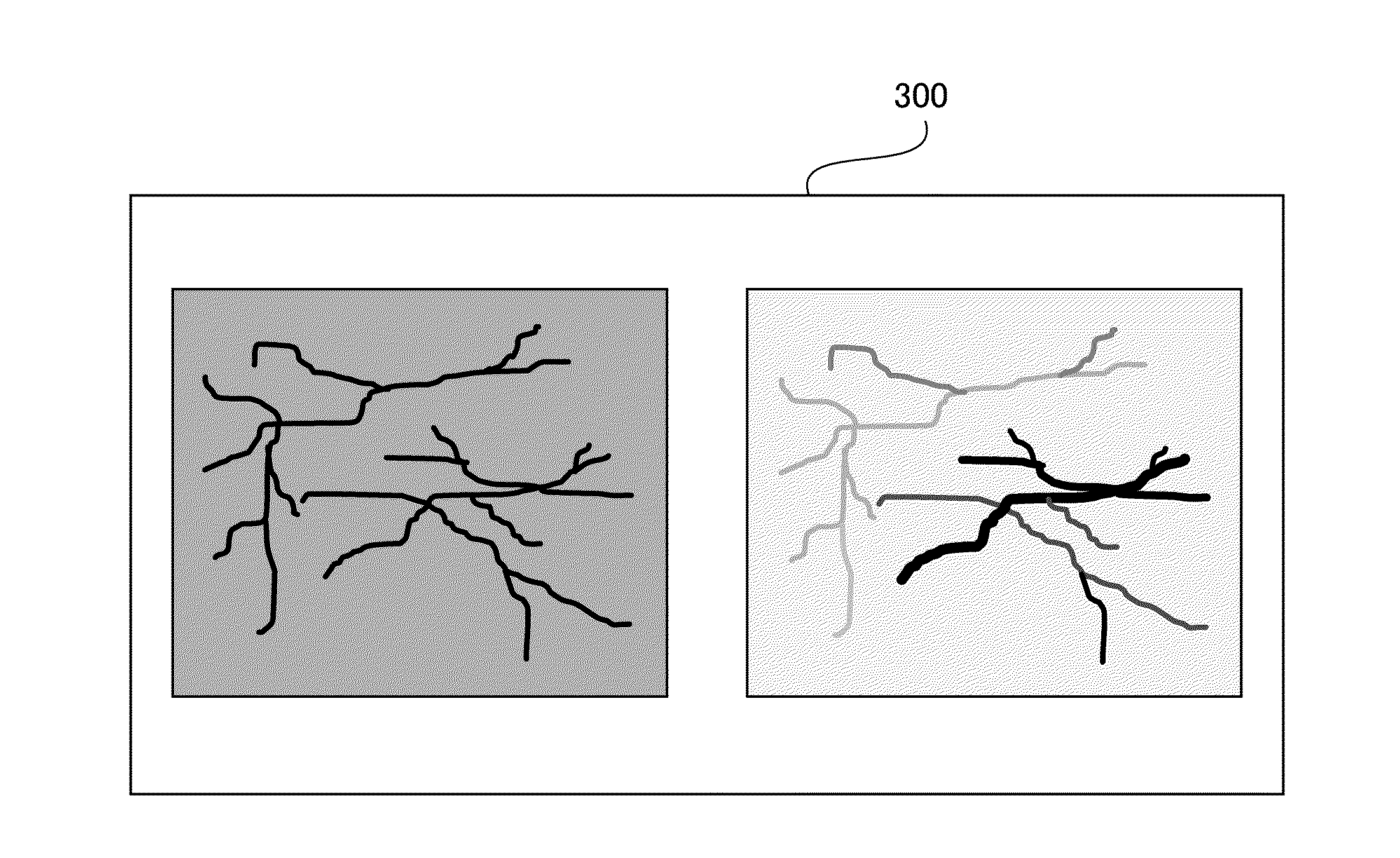
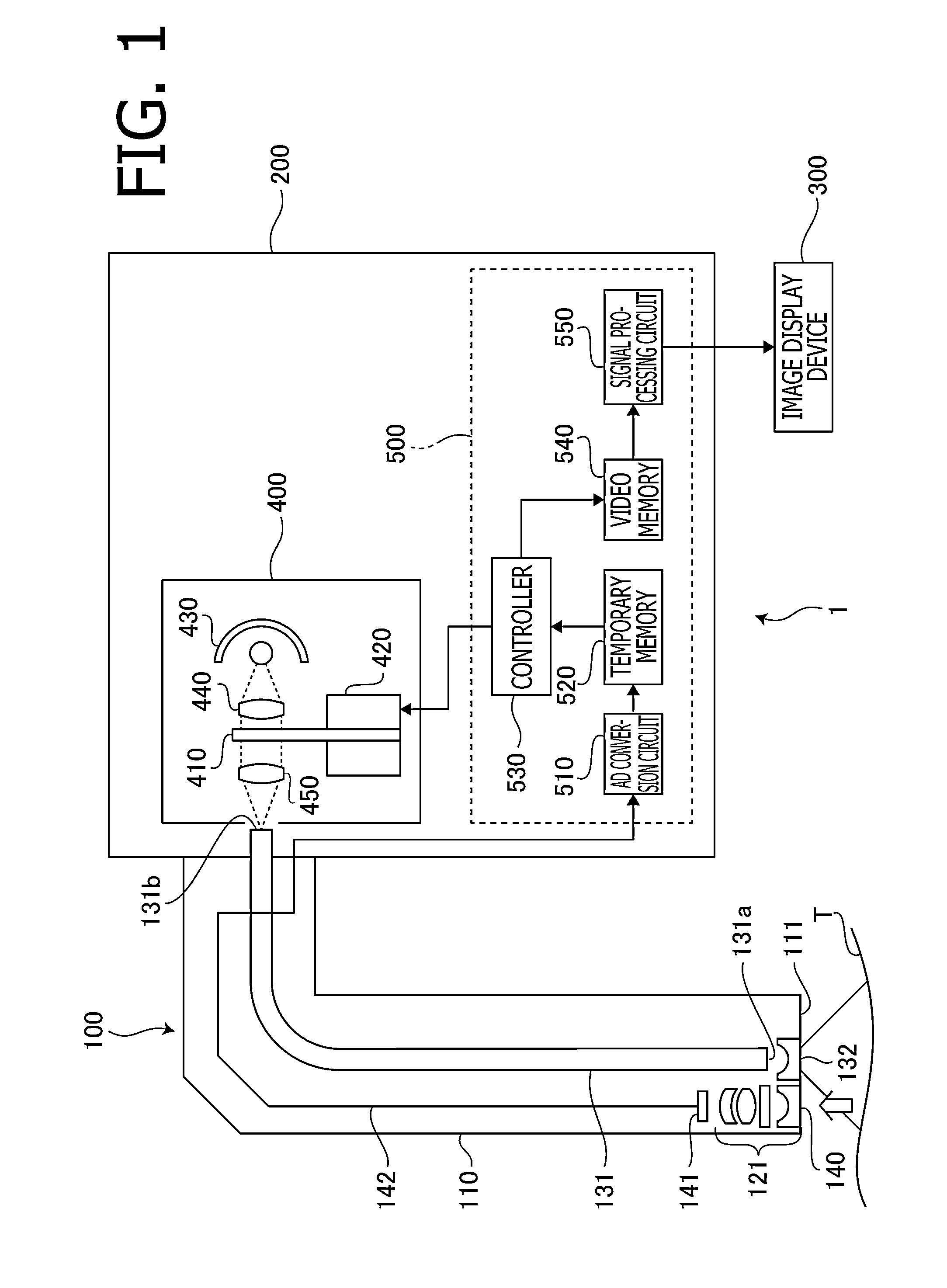
[0025]FIG. 1 is a block diagram of a diagnostic system 1 according to the embodiment of the invention. The diagnostic system 1 according to the embodiment is configured to generate an indicator image to be referred to by a medical doctor for diagnosing a disease of a digestive organ, such as a stomach or a rectum. The diagnostic system 1 includes an electronic endoscope 100, a processor 200 for an electronic endoscope and an image display device 300. In the processor 200 for an electronic endoscope, a light source unit 400 and an image processing unit 500 are accommodated.

[0026]The electronic endoscope 100 includes an insertion tube 110 to be inserted into a body cavity, and an objective optical system 121 is provided at a tip portion (an insertion tube tip portion) 111 of the insertion tube 110. An image of a living tissue T around the insertion tube tip portion...


PUM


Abstract

A diagnostic system comprises a spectral image pickup means that picks up a spectral image in a predetermined wavelength region in a body cavity and obtains spectral image data, an image processing means that obtains, from the spectral image data, an index-value for discriminating between a diseased portion and a healthy portion, and generates and outputs an indicator image based on the index-value, and a monitor on which the indicator image is displayed, wherein, for each pixel of the spectral image, the image processing means defines β obtained by a predetermined expression as the index-value, while using the spectral image data P1 at a first wavelength which is around a wavelength of 542 nm, the spectral image data P2 at a second wavelength which is around a wavelength of 558 nm and the spectral image data P3 at a third wavelength which is around a wavelength of 578 nm.

Description

TECHNICAL FIELD[0001]The present invention relates to a diagnostic system configured to display an image of a region that is highly likely to be a diseased portion in a living tissue.BACKGROUND ART[0002]Recently, an electronic endoscope having a function as a spectrometer has been proposed as described, for example, in Japanese Patent Provisional Publication No. JP2007-135989A. By using such an electronic endoscope, it is possible to obtain the spectral property (the distribution of light absorption property for each frequency) of a living tissue such as a mucous membrane of a digestive organ, e.g., a stomach or a rectum. It is known that the spectral property of a substance reflects information concerning the types or densities of components contained in the vicinity of a surface layer of a living tissue being an observation target, which is established in a field belonging to academic frameworks of analytical chemistry. It is also known in this field that the spectral property of ...

Claims


Application Information

Patent Timeline
IPC IPC(8): G06K9/00A61B8/08
CPCA61B8/48G06K9/0014A61B1/0638A61B1/0646A61B1/0005G06T7/0012G06T2207/10068A61B5/6871A61B5/6873G06T2207/10024G06T2207/30092G06T2207/30096G06V20/695A61B1/000094
Inventor CHIBA, TORU
Owner HOYA CORP
Why Eureka
Try Eureka