Method to operate an image-generating medical modality to avoid patient injury by a modality-generated electromagnetic field

Inactive Publication Date: 2013-10-03
SIEMENS HEATHCARE GMBH
View PDF1 Cites 3 Cited by
  • Summary
  • Abstract
  • Description
  • Claims
  • Application Information

AI Technical Summary

Benefits of technology

This patent text discusses a method for reducing harmful electromagnetic fields that are used to examine patients. The method takes into account the fact that the fields are potentially harmful and are not evenly distributed through the patient. Instead, there is a risk of injury depending on the patient's position in the field. The method checks if the patient's body extends to critical regions and adjusts the field accordingly to avoid high power in those areas. This process is automated, making it easier to operate the medical modality and reducing the likelihood of errors. The goal is not to achieve high resolutions or show details, but rather to emphasize contours and present without distortion. This allows for precise measurement of the patient's extent and the smallest clearance between them and the modality's wall.

Problems solved by technology

Consideration is given to the fact that electromagnetic fields that are used to examine patients are potentially harmful to the patient and are not homogeneous with regard to the spatial distribution.
The radiation exposure of the patient thus exhibits local differences, and in some regions an undesirably high radiated power is sometimes present that entails an impermissibly high risk of an injury to the patient.
At a given mean radiated power, the maximum local radiation exposure can thus be tolerable in one position but can already produce an injury to the patient in another, more critical position.

Method used


Image

  • Method to operate an image-generating medical modality to avoid patient injury by a modality-generated electromagnetic field
  • Method to operate an image-generating medical modality to avoid patient injury by a modality-generated electromagnetic field
  • Method to operate an image-generating medical modality to avoid patient injury by a modality-generated electromagnetic field

Examples


Embodiment Construction

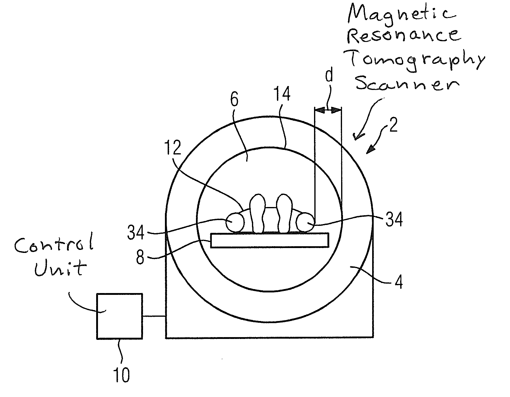
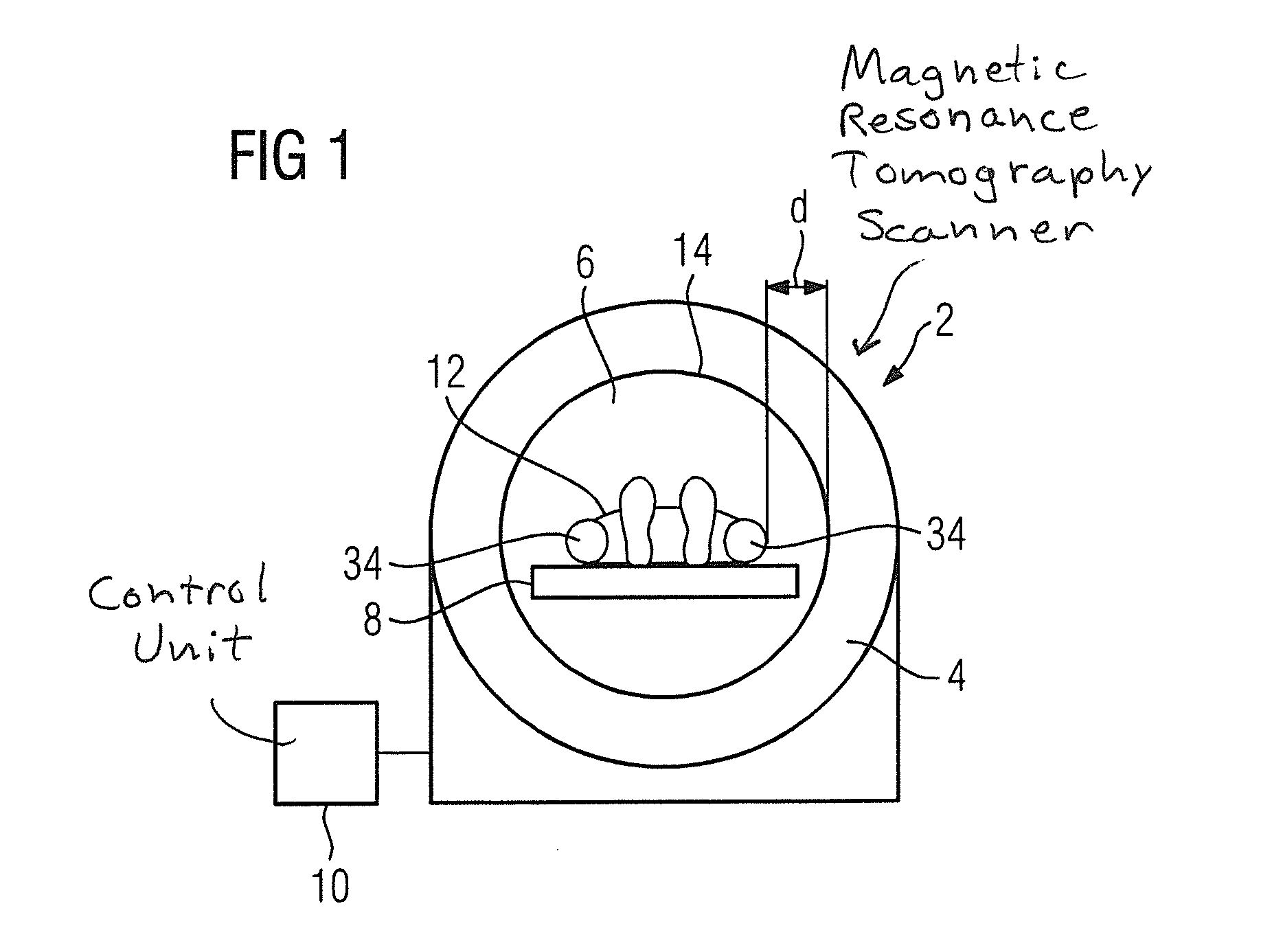
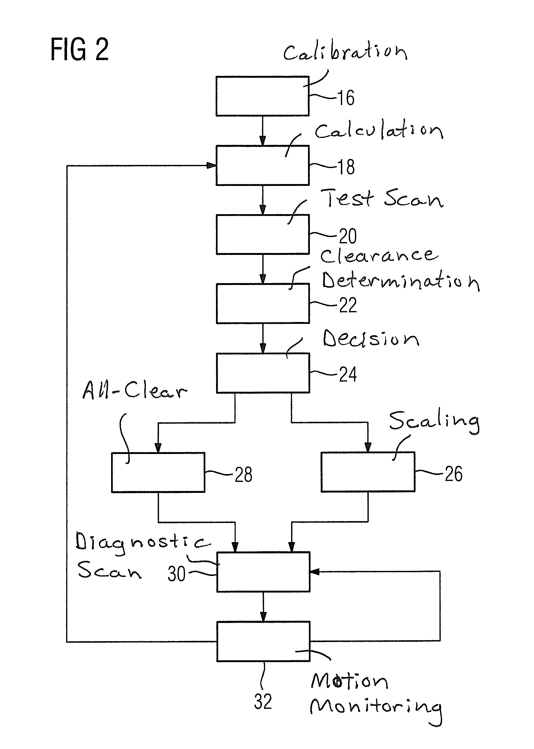
[0031]Parts corresponding to one another are respectively provided with the same reference characters in all figures.

[0032]For example, the method presented here is suitable for operation of a magnetic resonance tomography scanner 2 as it is schematically shown in FIG. 1. This includes a base unit 4 with a centrally arranged and cylindrical chamber (also called a tunnel 6 in the following), a patient table 8 and a control unit 10. Integrated into the base unit 4 is a coil arrangement (not shown in detail) that is designed according to a known principle for the generation of the required fields, thus a radio-frequency field, three low-frequency gradient fields and a static magnetic field. The individual modules / sub-coils of the coil arrangement are typically arranged around the tunnel with the radio-frequency coils inside the gradient coil coils, which in turn are inside the magnet that generates the static magnetic field. With the use of such a magnetic resonance tomography scanner ...


PUM


Abstract

In a method to operate an image-generating medical modality in order to avoid harm to a patient caused by an electromagnetic field, an adjustment process is implemented in which the electromagnetic field is radiated by the modality and preliminary image data are acquired using the electromagnetic field; and a clearance between the patient and a wall of the modality is determined from the image. The determined clearance forms the basis of a check process to determine an adaptation of the electromagnetic field for a diagnostic examination of the patient that follows. During the diagnostic examination, that also includes radiation of the electromagnetic field, movement monitoring of the patient takes place and the adjustment process is restarted as soon as a movement of the patient is registered.

Description

BACKGROUND OF THE INVENTION[0001]1. Field of the Invention[0002]The invention concerns a method to operate an image-generating medical modality (in particular a magnetic resonance tomograph) and a corresponding image-generating medical modality.[0003]2. Description of the Prior Art[0004]A magnetic resonance tomography (MRT) apparatus frequently has a tube-like stationary unit with which the electromagnetic fields required for an imaging method are generated during operation. Such a tube-like stationary unit has a tunnel-like and centrally arranged opening or recess in which the generated electromagnetic fields act during operation, and in which a subject to be examined (a patient, for example) is positioned for an examination by means of imaging methods.[0005]This tunnel-like recess is bounded by parts of the surface of the stationary unit that are designated herein as an inner tunnel wall. Located directly behind the inner tunnel wall are some of the assemblies of the stationary un...

Claims


Application Information

Patent Timeline
IPC IPC(8): G01R33/28
CPCG01R33/288G01R33/56572G01R33/56563G01R33/543
Inventor BLUMHAGEN, JAN OLEBRINKER, GERHARD
Owner SIEMENS HEATHCARE GMBH
Why Eureka
Try Eureka