
System for Motion Corrected MR Diffusion Imaging

a technology of diffusion imaging and motion correction, applied in the field of system for motion correction mr diffusion imaging, can solve the problems of bulk subject motion, erroneous calculation of diffusion parameters, signal loss in mr images,

Active Publication Date: 2013-07-25
SIEMENS HEALTHCARE GMBH +1
View PDF12 Cites 43 Cited by
  • Summary
  • Abstract
  • Description
  • Claims
  • Application Information

AI Technical Summary

Benefits of technology

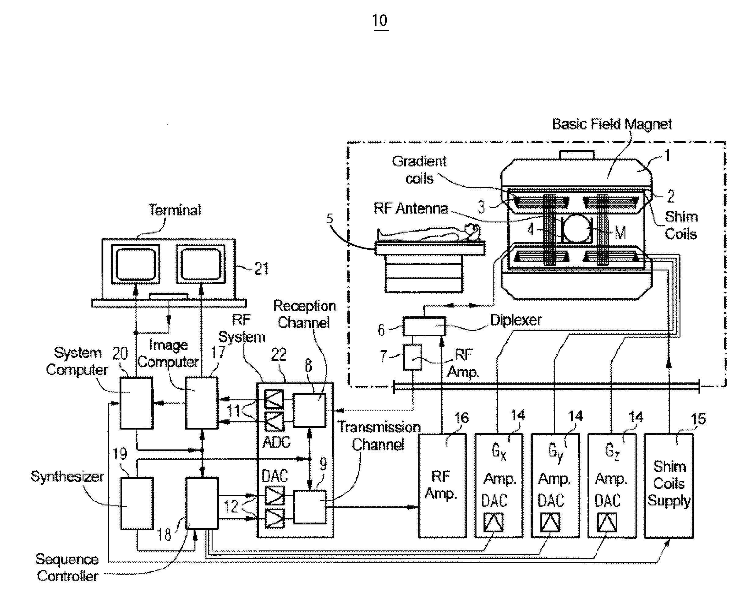
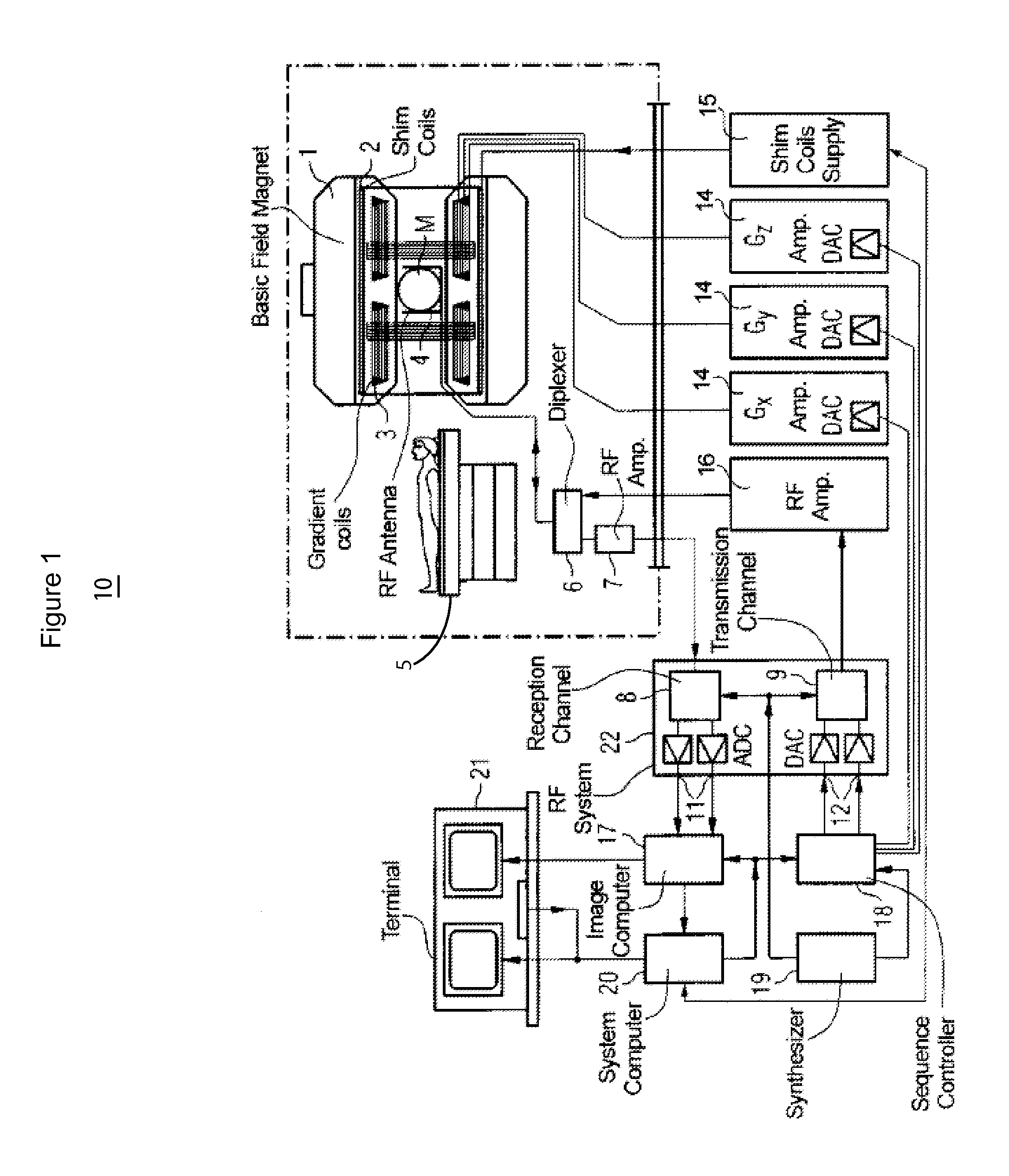
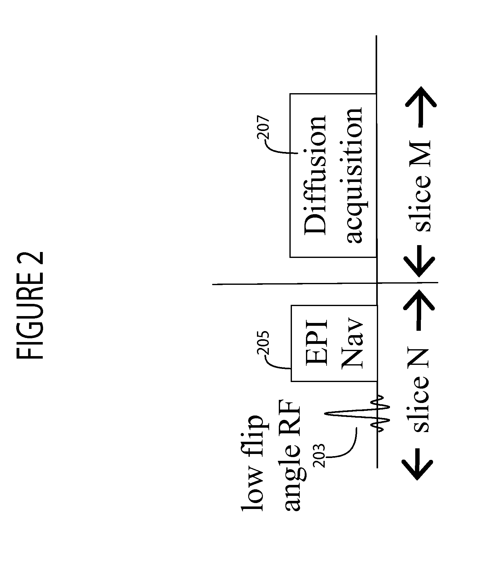
This patent describes a system for improving the motion correction data in diffusion MR imaging of an anatomical volume. The system uses a combination of RF signal generation and magnetic field gradient generation to acquire RF echo data and prepare for acquiring data representing a diffusion image slice. The system can acquire a 2D non-diffusion image and a diffusion image slice in a single direction through the volume, and can also provide a sequence for acquiring multiple diffusion image slices. The technical effect of this patent is to improve the accuracy and efficiency of diffusion MR imaging by accounting for motion during the acquisition process.

Problems solved by technology

In the presence of a magnetic field gradient, diffusion of water molecules leads to signal loss in MR images.
For such long scans, bulk subject motion is a problem.
In diffusion neuroimaging, problems occurring due to patient motion include, i) images acquired with different diffusion directions are misaligned, leading to erroneous calculation of diffusion parameters, and ii) images being acquired in a diffusion direction when motion occurs are susceptible to MR signal dropouts.
Multiple known systems, both retrospective and prospective, attempt to address this problem with limited success.

Method used


Image

  • System for Motion Corrected MR Diffusion Imaging
  • System for Motion Corrected MR Diffusion Imaging
  • System for Motion Corrected MR Diffusion Imaging

Examples


Embodiment Construction

[0018]A system in one embodiment provides a rigid body prospective motion correction method for diffusion imaging using interleaved and integrated non-diffusion encoded low resolution Echo planar imaging (EPI) images as navigators. A navigator image determines position of an anatomical structure e.g. the diaphragm before image data acquisition of a desired region of interest. Navigator images are used to identify respiratory and other movement of the patient for synchronizing image data acquisition so that movement induced image blurring is minimized, for example. The system provides prospective motion correction for multi-slice single shot diffusion weighted EPI. The system provides prospective motion correction for 2D multislice diffusion imaging, by using non-diffusion encoded low resolution single shot EPI images as motion navigator (EPI Navigator images) during a diffusion scan. The system provides a prospective motion correction method for multi-slice single shot diffusion wei...


PUM


Abstract

A system determines motion correction data for use in diffusion MR imaging using an RF signal generator and magnetic field gradient generator which sequentially acquire in a single first direction through a volume, first and second slice sets individually comprising multiple individual diffusion image slices. The first set of slices and the second set of slices are spatially interleaved within the volume, by providing in acquiring the second slice set, a low flip angle RF pulse successively followed by a non-diffusion image data readout magnetic field gradient for acquisition of data representing a two dimensional (2D) non-diffusion image used for motion detection of the first slice set successively followed by, a first diffusion imaging RF pulse followed by a first diffusion imaging phase encoding magnetic field gradient for preparation for acquiring data representing a diffusion image slice of the second slice set.

Description

CROSS-REFERENCE TO RELATED APPLICATIONS[0001]This application is a non-provisional of, and claims priority to, U.S. provisional patent application Ser. No. 61 / 589,969 by H. Bhat et al. filed on 24 Jan., 2012, incorporated herein by reference in its entirety.FIELD OF THE INVENTION[0002]This invention concerns a system for determining motion correction data for use in diffusion MR imaging of an anatomical volume.BACKGROUND OF THE INVENTION[0003]Diffusion MRI is a magnetic resonance imaging (MRI) method that produces in vivo images of biological tissues weighted with the local microstructural characteristics of water diffusion and is capable of showing connections between brain regions. In the presence of a magnetic field gradient, diffusion of water molecules leads to signal loss in MR images. The degree of signal loss depends on the characteristics of the diffusion, which in turn depends on tissue properties like structure, surrounding environment, physical state and pathology. The u...

Claims


Application Information

Patent Timeline
Patent Type & Authority Applications(United States)
IPC IPC(8): G01R33/44
CPCA61B5/055G01R33/5676G01R33/56341
Inventor BHAT, HIMANSHUVAN DER KOUWE, ANDRE JAN WILLEMTISDALL, MATTHEW DYLANHEBERLEIN, KEITH AARON
Owner SIEMENS HEALTHCARE GMBH
Learn More