Methods for diagnosing solid tumors
a solid tumor and method technology, applied in the field of genetics and cellular and molecular biology, can solve the problems of inability to achieve the same progress in diagnosis or prognosis of disease, or the expansion of knowledg
- Summary
- Abstract
- Description
- Claims
- Application Information
AI Technical Summary
Benefits of technology
Problems solved by technology
Method used
Image
Examples
example 1
General Methology for Cell Analysis
[0321]Cell thawing, ficoll density gradient separation, and live / dead staining: Cells are thawed in a 37° C. water bath in cryovials. Once the cells are thawed, 1 mL of pre-warmed thaw buffer (RPMI+60% FBS) is added dropwise to the cryovials and then the entire contents of the cryovials are transferred to a 15 mL conical tube. The volume of each sample is brought up to 10 mL by adding the appropriate volume of thaw buffer. The 15 mL tubes are then capped and inverted 3 times.
[0322]A ficoll density gradient separation is then performed by underlaying 5 mL of ambient temperature ficoll using a Pasteur pipette on the samples. Next, the tubes are centrifuged at 400×g for 30 minutes at room temperature, the “buffy coat” aspirated, and the mononuclear cell layer transferred to a new 15 mL conical tube containing 9 mL thaw buffer. The cell layers are centrifuged at 400×g for 5 minutes, the liquid aspirated, the cell pellet gently resuspended. Subsequently...
example 2
SCNP Differs Between Cleaved-PARP+ and Cleaved-PARRneg Cell Populations
[0331]Intracellular network responses of AML patient samples were modulated by SCF and analyzed using flow-cytometry based Single Cell Network Profiling (SCNP). The protocol was similar to the protocols listed above and in U.S. Ser. Nos. 61 / 350,864 and 12 / 460,029. For each sample, the CD34+ cell population was further gated into cleaved-PARP+and cleaved-PARPneg cell populations using software such as Flowjo, DIVA (available from Becton Dickinson), and / or Winlist (available from Verity Software). The Mean Fluorescence Intensity (MFI) of various signaling nodes (pAKT, pS6, and pERK) obtained by flow-cytometry based SCNP was examined in as function of cleaved-PARP gating.
[0332]Gating scheme 1 does not incorporate c-PARP as part of the analysis. Gating scheme 2 excludes all c-PARP+ (apoptotic) cells. Gating scheme 3 incorporates only c-PARP+ cells. The utility of incorporating c-PARP in the gating analysis allows one...
example 3
Ananlysis of Cells in the Bladder Cancer Wash Samples
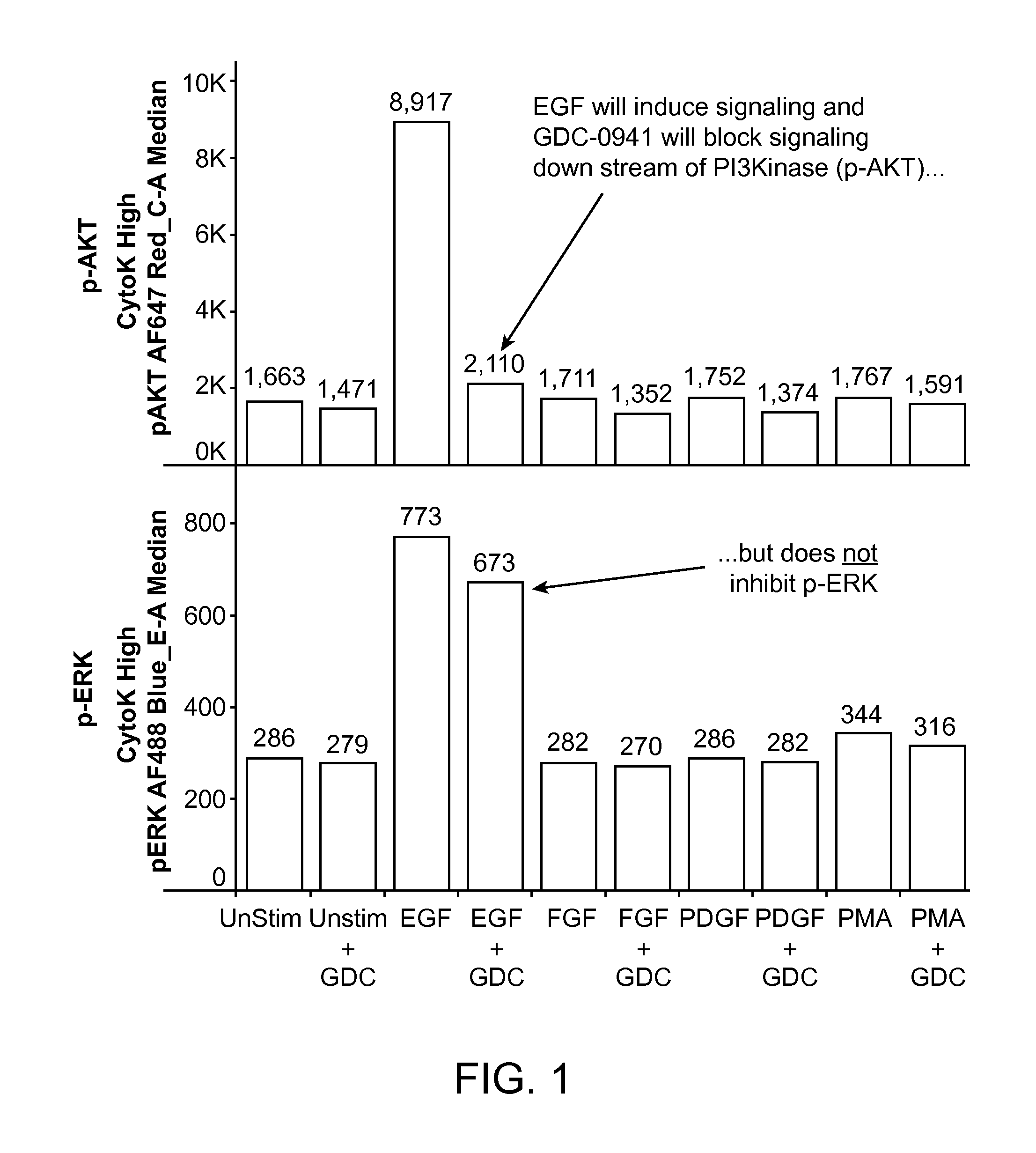
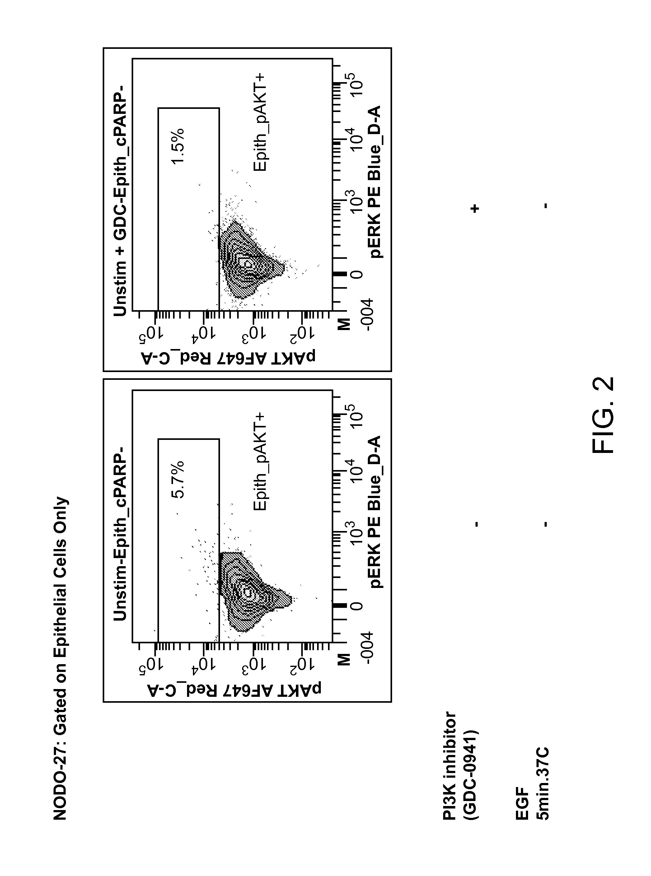
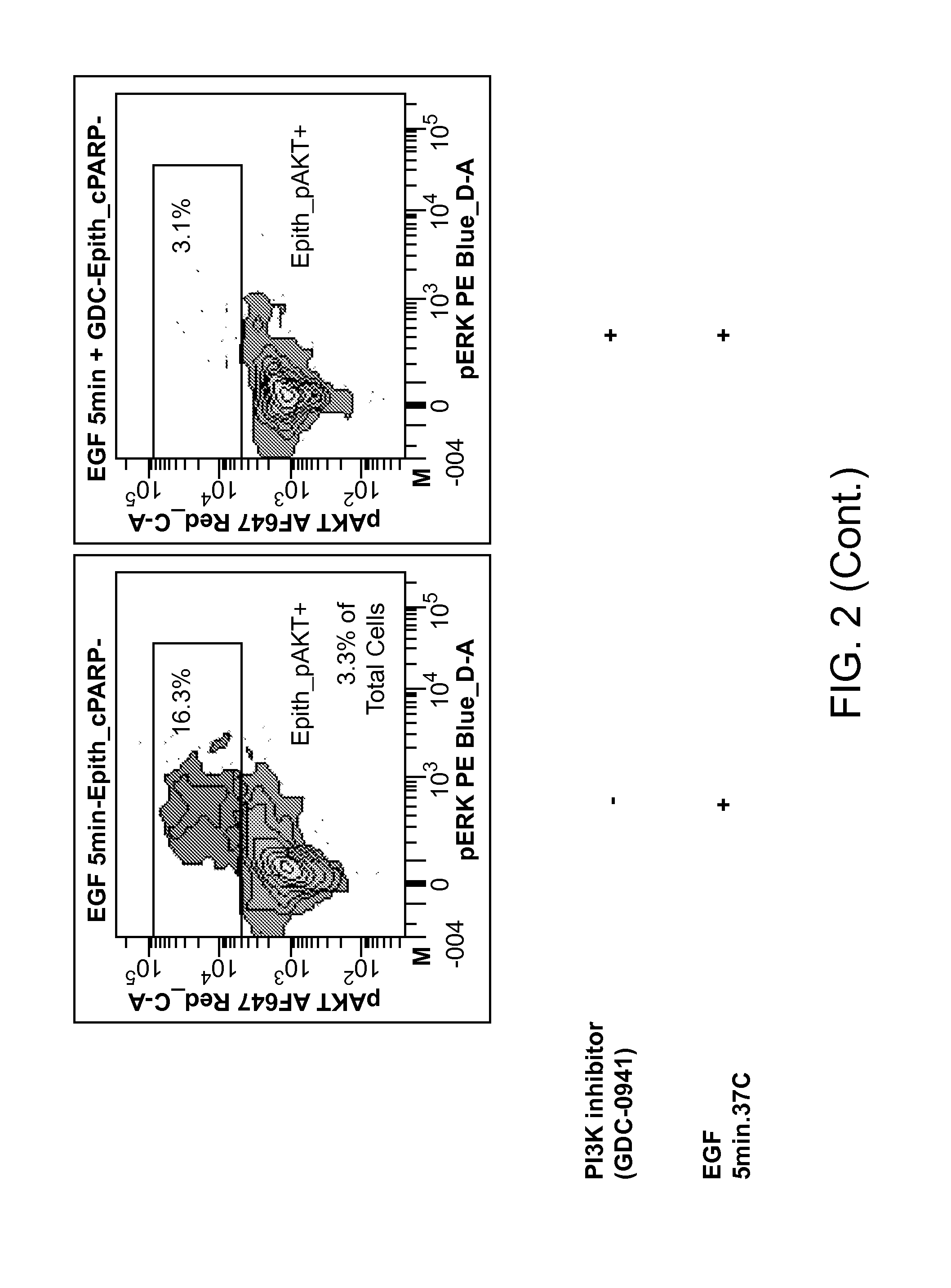
[0335]Bladder washes from non-cancer (NC) patients (pts) (n=8) and confirmed / suspected bladder cancer (BC) pts (n=20) were collected using standard medical practice and shipped overnight on ice for processing within 24 hours. Antibodies against CD45, Cytokeratin (CK), and EpCAM were used to differentiate epithelial cells from leukocytes, while measurements of DNA aneuploidy (DAPI stain) allowed for distinction between tumor and normal epithelial cells. Signaling activity in the PI3K and MAP Kinase pathways was assessed by measuring intracellular levels of p-AKT and p-ERK both at baseline and in response to pathway modulation. A process similar to that shown in Example 1 was used.
[0336]Upon delivery, cells were pelleted, counted, rested for 1 hr at 37° C., followed by a 5 minute stimulation with EGF or vehicle+ / −GDC-0941 200 nM added 1 hr prior to EGF addition to inhibit PI3K signaling After cell fixation and permeabilization, samp...
PUM
| Property | Measurement | Unit |
|---|---|---|
| pore sizes | aaaaa | aaaaa |
| mass spectrometer | aaaaa | aaaaa |
| Multi Volume Treatise | aaaaa | aaaaa |
Abstract
Description
Claims
Application Information
 - R&D
- Intellectual Property
- Life Sciences
- Materials
- Tech Scout
- Unparalleled Data Quality
- Higher Quality Content
- 60% Fewer Hallucinations
Browse by: Latest US Patents, China's latest patents, Technical Efficacy Thesaurus, Application Domain, Technology Topic, Popular Technical Reports.
© 2025 PatSnap. All rights reserved.Legal|Privacy policy|Modern Slavery Act Transparency Statement|Sitemap|About US| Contact US: help@patsnap.com


